RadQBoard › Musculoskeletal › Chapter 1
Bones, joints, osteoarthritis and rheumatoid arthritis
This chapter starts with how bones and synovial joints are built, then follows osteoarthritis joint by joint, from erosive hand osteoarthritis to the way the femoral head migrates in the hip, and DISH. The second half covers rheumatoid arthritis: the synovial disease, its pattern in the hands and feet, even narrowing of large joints, and instability at C1–C2.
Read all 10 pages and answer the chapter's 32 questions in the app. Free to read, no account needed.
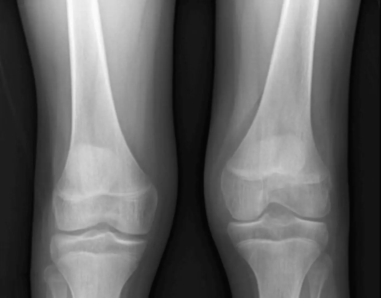
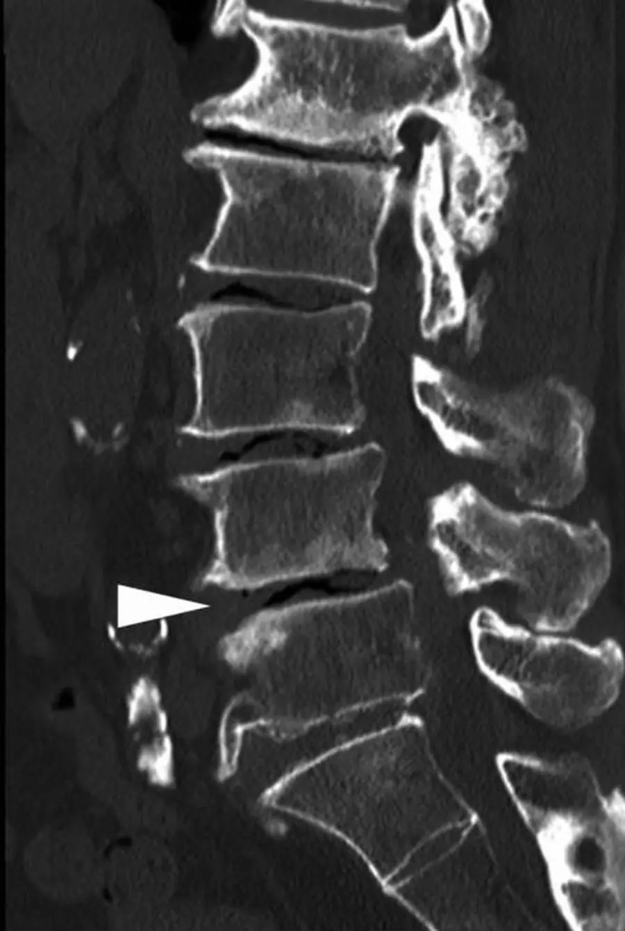
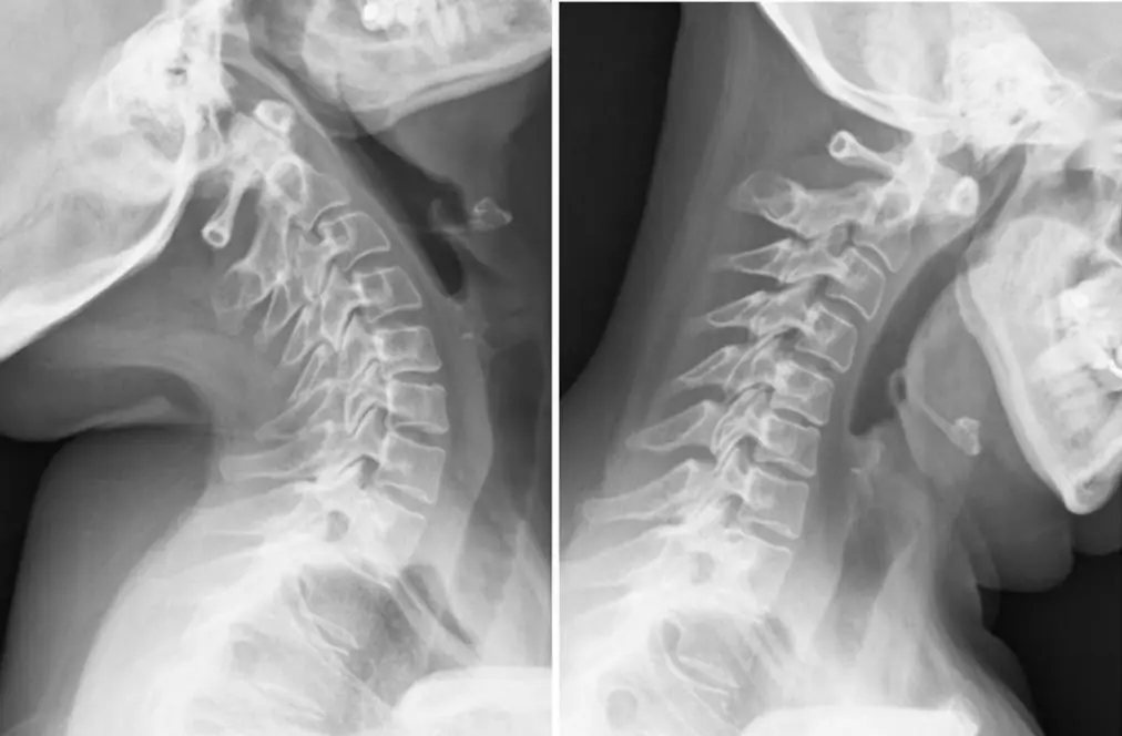
What this chapter covers
Bones and joints: the basicsFree
Know the parts of a long bone and a synovial joint, and every arthritis becomes a question of where the damage starts.
Osteoarthritis: the diseaseFree
Osteoarthritis wears cartilage away where the load falls, and the bone answers with osteophytes, sclerosis and cysts.
Osteoarthritis of the hand and erosive OAFree
In the hand, OA lives at the DIP joints, the thumb base and the PIP joints, and leaves the MCP joints alone.
Osteoarthritis of the knee, shoulder and footFree
Weight-bearing views tell the truth about the knee; the Grashey view tells it about the shoulder.
Osteoarthritis of the hipFree
The direction the femoral head moves tells you the arthritis: up and out in OA, straight in along the axis in rheumatoid arthritis.
Degenerative spine and DISHFree
The disc degenerates; the facets get osteoarthritis. They are two different processes in two different kinds of joint.
Rheumatoid arthritis: the diseaseFree
Rheumatoid arthritis is an attack on the synovium; the bone loss is collateral damage, starting where synovium touches bare bone.
Rheumatoid arthritis of the hand and wristFree
MCP, PIP and wrist, both sides, with the DIP joints spared: the rheumatoid hand.
Rheumatoid arthritis of the foot, hip and kneeFree
In the foot RA can be the first sign; in the hip and knee it narrows the whole joint evenly.
Rheumatoid arthritis of the cervical spineFree
RA erodes and loosens the neck without building any bone; the danger point is C1–C2.
Further reading
- Roemer FW, Demehri S, Omoumi P, et al. State of the Art: Imaging of Osteoarthritis—Revisited 2020. Radiology. 2020;296(1):5-21. doi:10.1148/radiol.2020192498
- Martel W, Stuck KJ, Dworin AM, et al. Erosive osteoarthritis and psoriatic arthritis: a radiologic comparison in the hand, wrist, and foot. American Journal of Roentgenology. 1980;134(1):125-135. doi:10.2214/ajr.134.1.125
- Modic MT, Ross JS. Lumbar Degenerative Disk Disease. Radiology. 2007;245(1):43-61. doi:10.1148/radiol.2451051706
- Resnick D, Niwayama G. Radiographic and Pathologic Features of Spinal Involvement in Diffuse Idiopathic Skeletal Hyperostosis (DISH). Radiology. 1976;119(3):559-568. doi:10.1148/119.3.559
- Sommer O, Kladosek A, Weiler V, et al. Rheumatoid Arthritis: A Practical Guide to State-of-the-Art Imaging, Image Interpretation, and Clinical Implications. RadioGraphics. 2005;25(2):381-398. doi:10.1148/rg.252045111
- Martínez de Vega L, Baker J, Suevos-Ballesteros C, et al. A Radiologic Walk through Rheumatoid Arthritis. RadioGraphics. 2026;46(3):e250059. doi:10.1148/rg.250059
- Morimoto LR, Kase DT, Esmanhotto PG, et al. Imaging Assessment of Nontraumatic Pathologic Conditions at the Craniovertebral Junction: A Comprehensive Review. RadioGraphics. 2024;44(5):e230137. doi:10.1148/rg.230137
Read all 10 pages and answer the chapter's 32 questions in the app. Free to read, no account needed.