RadQBoard › Musculoskeletal › Chapter 1 › Bones and joints: the basics
Bones and joints: the basics
Know the parts of a long bone and a synovial joint, and every arthritis becomes a question of where the damage starts.
What bone is made of
Bone has two structural forms, and both give the rigid frame that muscles move:
- Cortical (compact) bone is the dense outer shell. Its osteons are tightly packed, each with a central Haversian canal carrying vessels and nerves, and osteocytes sit in lacunae linked by canaliculi.
- Trabecular (cancellous) bone fills the center as a lattice of struts with marrow between them. Its osteons are loosely arranged and lack a central canal, but share the layered structure and osteocytes of cortical bone.
Trabecular bone turns over faster, so it shows osteoporosis, marrow disease and metastases earlier than cortex.
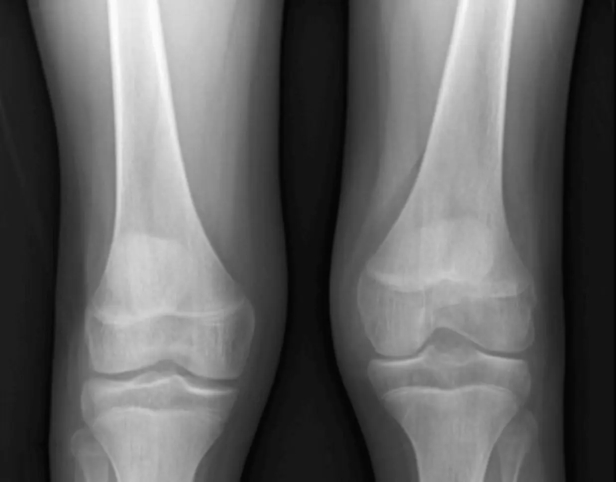
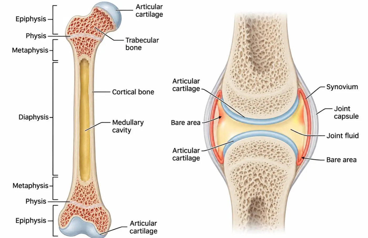
Parts of a long bone
| Part | Where | Why it matters |
|---|---|---|
| Epiphysis | The end of the bone, inside the joint | Articular cartilage; site of chondroblastoma and giant cell tumor (after fusion) |
| Physis | The growth plate | Growth; fractures in children (Salter-Harris) |
| Metaphysis | The flared part next to the physis | Rich blood supply; the most common site of primary bone tumors and of hematogenous osteomyelitis in children |
| Diaphysis | The shaft | Round cell tumors, fibrous lesions, stress fractures |
The synovial joint
- Articular cartilage (hyaline) covers the bone ends. It has no nerves or vessels and cannot be seen on radiographs: its thickness is the "joint space".
- Synovium lines the capsule and makes synovial fluid.
- Capsule and ligaments hold the joint together.
- The bare area is bone inside the capsule that is not covered by cartilage. Inflamed synovium touches it directly, so it is where the first erosions of rheumatoid arthritis appear (marginal erosions).
Other joints are cartilaginous (the disc-vertebral body junction, the pubic symphysis) or fibrous (skull sutures). Osteoarthritis affects synovial joints; the disc degenerates by a different process.
Arthritis: the categories
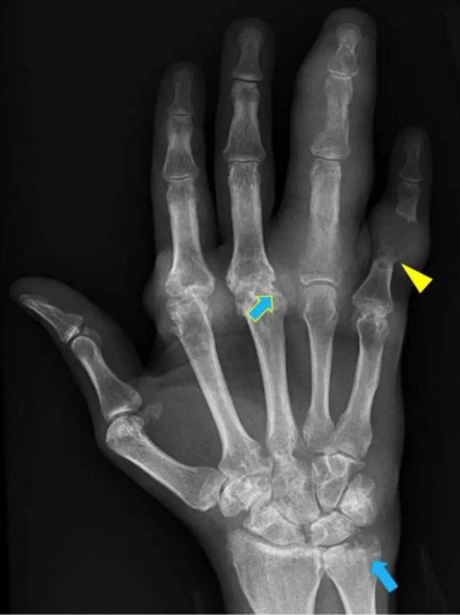
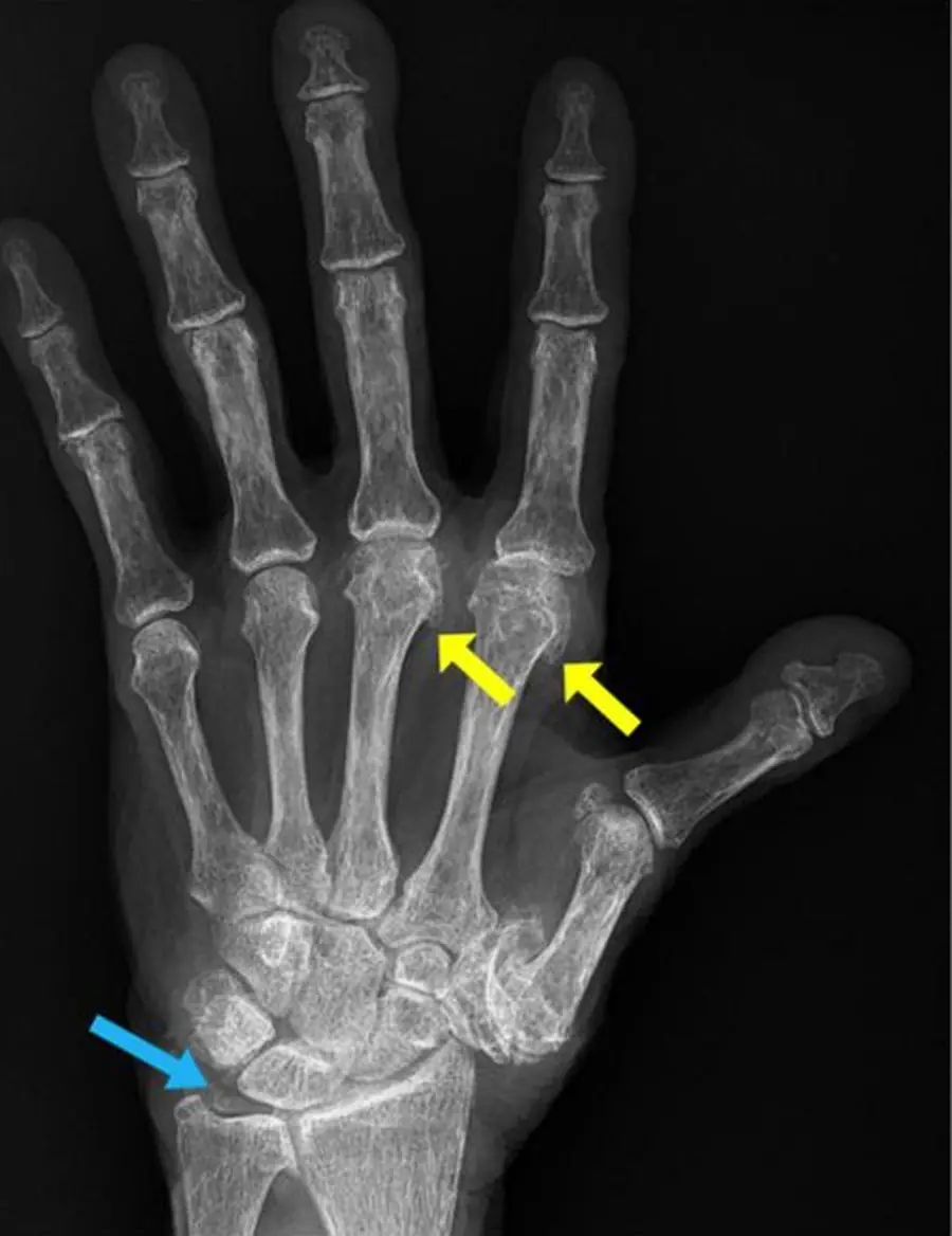
The hallmark of every arthritis is cartilage loss, seen as joint-space narrowing. The pattern of everything else separates the types:
- Degenerative: osteoarthritis.
- Inflammatory: rheumatoid arthritis, the spondyloarthropathies, juvenile idiopathic arthritis.
- Crystal deposition: gout, calcium pyrophosphate (CPPD), hydroxyapatite.
- Hematologic: hemophilia.
- Metabolic: hemochromatosis, ochronosis, acromegaly.
- Infection: septic arthritis, a separate emergency.
How to read an arthritis radiograph
Go through five questions, then match the pattern:
- Alignment: subluxation, deviation, collapse.
- Bone density: normal, or osteopenic around the joints.
- Cartilage space: narrowed uniformly or in one compartment.
- Distribution: which joints, one side or both, symmetric or not.
- Soft tissues: swelling, tophi, calcification.
Then look for erosions (where, what shape) and new bone (osteophytes, periostitis, enthesophytes).
| Pattern | Osteoarthritis | Rheumatoid arthritis | Spondyloarthropathy | Gout | CPPD |
|---|---|---|---|---|---|
| Joint space | Non-uniform narrowing | Uniform narrowing | Narrowed, may fuse | Preserved until late | Narrowed in unusual joints |
| Bone density | Normal | Periarticular osteopenia | Normal | Normal | Normal |
| Erosions | None (central in erosive OA) | Marginal | Marginal, with new bone | Punched out, overhanging edge | None; large cysts |
| New bone | Osteophytes, sclerosis | Little | Periostitis, enthesophytes | None | Osteophytes |
| Tell-tale | DIP, knee, hip, 1st CMC | MCP, PIP, wrist, MTP | SI joints, spine, heel | 1st MTP, tophi | Chondrocalcinosis |
- Judge knee joint space on weight-bearing views; supine views can hide cartilage loss.
Further reading
- Roemer FW, Demehri S, Omoumi P, et al. State of the Art: Imaging of Osteoarthritis—Revisited 2020. Radiology. 2020;296(1):5-21. doi:10.1148/radiol.2020192498
Practice this in RadQBoard. Chapter 1 has 32 board-style questions with full explanations. Free to read, no account needed.
Written in original words by the RadQBoard team. Images are reused under open licenses, with the source, license and any changes under each one.