RadQBoard › Sample questions
Sample radiology board questions
Ten questions from the RadQBoard question bank, one from each of ten musculoskeletal chapters. Each is built around a real, openly licensed image. Answer first, then open the explanation.
1. CT of the shin in a young woman with night pain
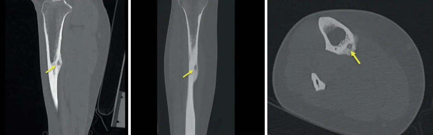
A 19-year-old woman has had aching pain in her left shin for 4 months. The pain is worst at night and goes away within an hour of taking ibuprofen. Sagittal, coronal and axial CT images of the tibia are shown. What is the most likely diagnosis?
- Brodie abscess
- Stress fracture
- Osteoid osteoma
- Osteoblastoma
- Chronic osteomyelitis with a sequestrum
Show the answer
Answer: C. Osteoid osteoma
CT shows a small, round lucent nidus with a central dot of mineralization inside a thickened, sclerotic cortex. In a young patient with night pain relieved by NSAIDs, this is osteoid osteoma. The nidus makes prostaglandins, which explains both the pain pattern and the response to aspirin or ibuprofen.
Key point: A small lucent nidus with a central calcified dot inside thick cortical sclerosis, in a young patient with night pain relieved by NSAIDs, is osteoid osteoma; thin-section CT is the best test to find it.
Topic: Osteoid osteoma. From Chapter 4: Bone-forming and cartilage-forming tumors. Practice the full block in RadQBoard.
2. Whole-body bone scan in an older man with back pain
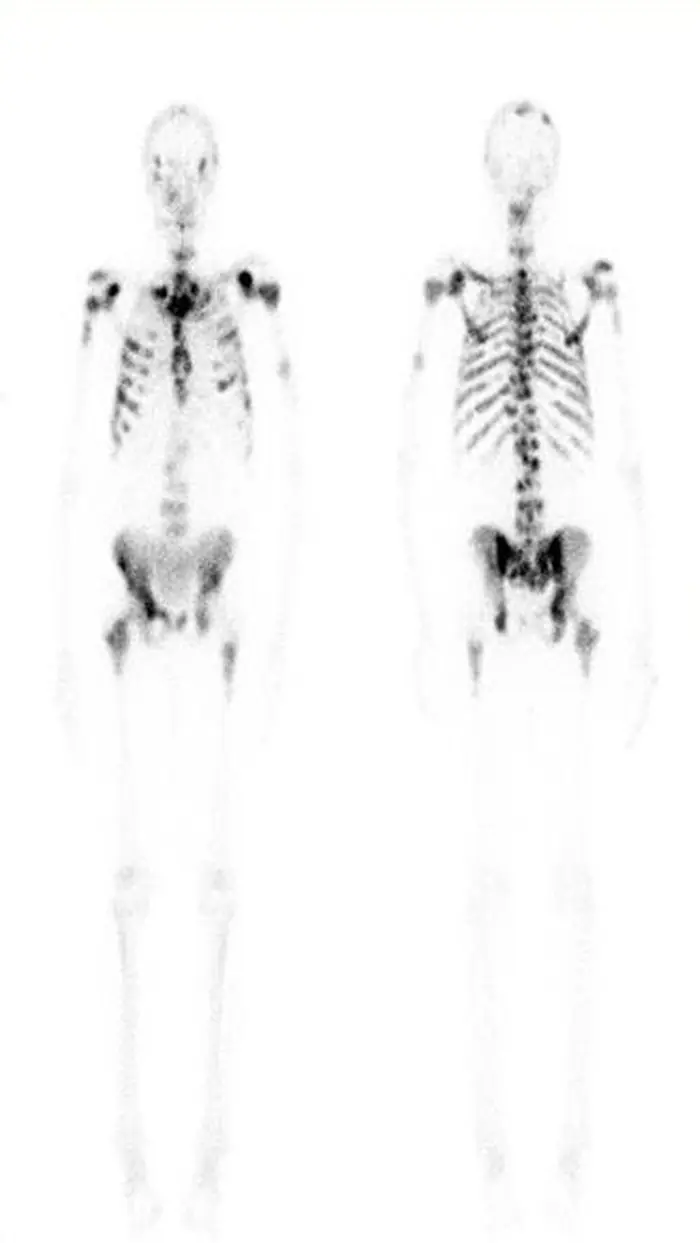
A 72-year-old man has back pain and weight loss. Anterior and posterior images from a Tc-99m MDP whole-body bone scan are shown. What is the most likely diagnosis?
- Metastatic prostate cancer
- Multiple myeloma
- Paget disease
- Rib fractures from a fall
- Renal osteodystrophy
Show the answer
Answer: A. Metastatic prostate cancer
Many randomly scattered foci of uptake of varying size in the spine, ribs, pelvis and shoulders, the red-marrow skeleton, in an older man are typical of blastic bone metastases, most often from prostate cancer.
Key point: Multiple randomly scattered foci of uptake in the axial skeleton of an older patient mean metastases; myeloma and aggressive lytic metastases can be cold on bone scan.
Topic: Bone metastases on bone scan. From Chapter 6: Bone cysts, metastases, tumor mimics and soft-tissue tumors. Practice the full block in RadQBoard.
3. MRI of a mass above the clavicle
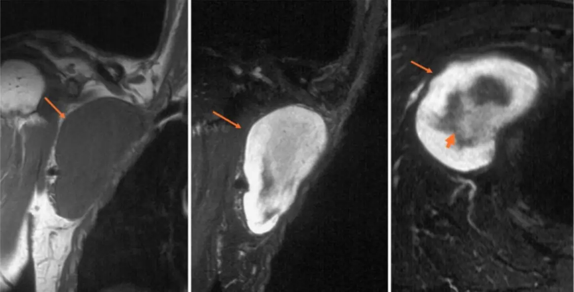
A 67-year-old man has a mass above his clavicle. Coronal T1-weighted, coronal fat-suppressed T2-weighted and axial fat-suppressed T2-weighted MRI images are shown. What is the appearance of the mass on the axial T2-weighted image (thick arrow) called?
- Target sign
- Split fat sign
- Fascicular sign
- Triple sign
- Bowl of grapes sign
Show the answer
Answer: A. Target sign
On the axial fat-suppressed T2 image, the center of the mass is darker than its bright rim: the target sign. It reflects central fibrous tissue and peripheral myxoid tissue and is typical of a nerve sheath tumor, especially neurofibroma.
Key point: The target sign, a dark center and bright rim on T2, suggests a benign nerve sheath tumor, especially neurofibroma, but does not prove it is benign.
Topic: Target sign. From Chapter 7: Nerve and other soft-tissue tumors, joint and bone infection. Practice the full block in RadQBoard.
4. Hand radiograph in a woman with kidney stones
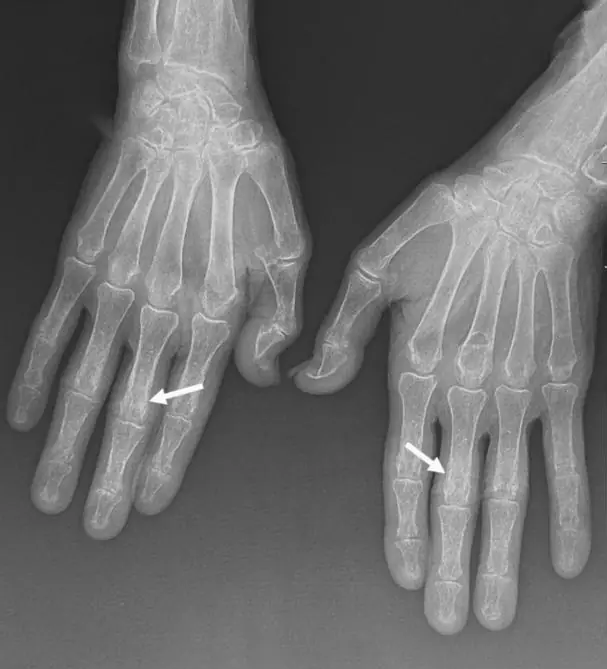
A 45-year-old woman has bone pain, kidney stones and a high serum calcium. A radiograph of both hands is shown. What is the most likely diagnosis?
- Scleroderma
- Rheumatoid arthritis
- Psoriatic arthritis
- Hyperparathyroidism
- Frostbite injury
Show the answer
Answer: D. Hyperparathyroidism
The cortical margins of the middle phalanges are lacy and eroded on both the radial and ulnar sides. Subperiosteal bone resorption, classically first on the radial side of the middle phalanges of the index and middle fingers, in a patient with hypercalcemia and stones, is hyperparathyroidism.
Key point: The most specific radiographic sign of hyperparathyroidism is subperiosteal resorption along the radial side of the middle phalanges of the index and middle fingers.
Topic: Hyperparathyroidism: subperiosteal resorption. From Chapter 8: Imaging infection and metabolic bone disease. Practice the full block in RadQBoard.
5. Spine radiographs in a teenager with anemia
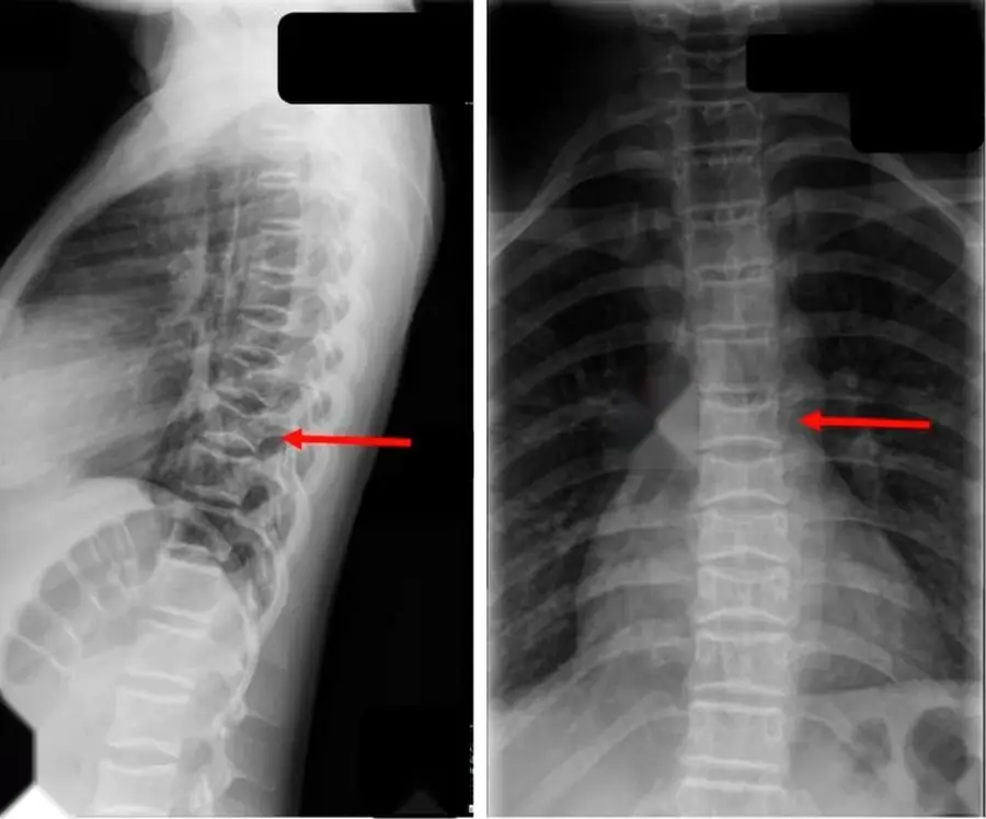
A 14-year-old with chronic anemia has had several painful crises requiring hospital care. Lateral and frontal radiographs of the thoracolumbar spine are shown. What is the most likely underlying condition?
- Osteoporosis
- Scheuermann disease
- Sickle cell disease
- Osteogenesis imperfecta
- Renal osteodystrophy
Show the answer
Answer: C. Sickle cell disease
The radiographs show square, flat-bottomed depressions of the central endplates, while the periphery of each endplate keeps its height: H-shaped vertebrae from infarction of the central growth zone. In a teenager with anemia and pain crises, this is sickle cell disease.
Key point: H-shaped vertebrae (square central endplate depressions) mean central endplate infarction: sickle cell disease with a small spleen, Gaucher disease with a large one.
Topic: Sickle cell disease: H-shaped vertebrae. From Chapter 9: Blood disorders of bone, lesion differentials, fractures and the forefoot. Practice the full block in RadQBoard.
6. Foot radiographs after a twisting fall
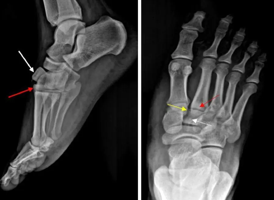
A 34-year-old man's foot was twisted under him when he fell down stairs with his foot pointed. Lateral and frontal radiographs of the foot are shown. Which joint is injured?
- Midtarsal (Chopart) joint
- Tarsometatarsal (Lisfranc) joint
- Subtalar joint
- Ankle (tibiotalar) joint
- First metatarsophalangeal joint
Show the answer
Answer: B. Tarsometatarsal (Lisfranc) joint
The lateral view shows dorsal displacement of the intermediate cuneiform and second metatarsal base, breaking the smooth dorsal outline of the tarsometatarsal joint, and the frontal view shows the second metatarsal base out of line with a fleck of bone beside it. This is a Lisfranc (tarsometatarsal) fracture-dislocation.
Key point: Lisfranc injury: the second metatarsal base slips out of line with the middle cuneiform, often with a dorsal step-off on the lateral view and a fleck of bone between the first two metatarsal bases.
Topic: Lisfranc fracture-dislocation. From Chapter 10: Midfoot, hindfoot and ankle. Practice the full block in RadQBoard.
7. Knee MRI in a man who can't straighten his knee
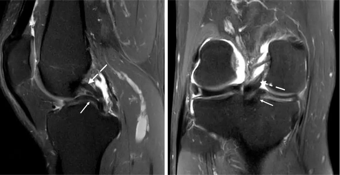
A 34-year-old man cannot fully straighten his knee after a twisting injury. Sagittal and coronal fat-suppressed MRI images are shown; the dashed arrow marks the native PCL. What does the structure at the solid arrow most likely indicate?
- A torn PCL with two stumps
- A ligament of Humphry
- A displaced medial bucket-handle fragment
- A displaced flap from a lateral meniscal horizontal tear
- A cruciate ligament ganglion
Show the answer
Answer: C. A displaced medial bucket-handle fragment
Just below and parallel to the native PCL lies a dark band, the displaced meniscal fragment, which looks like a second, smaller PCL. The double PCL sign indicates a bucket-handle tear, typically of the medial meniscus with an intact ACL, which prevents the fragment from flipping further forward.
Key point: Double PCL sign: a bucket-handle fragment lying in the notch under and parallel to the PCL, usually from the medial meniscus with an intact ACL; double delta (double anterior horn) suggests a lateral meniscal tear or a medial tear with a torn ACL.
Topic: Double PCL sign. From Chapter 11: Ankle fractures, ankle tendons and the knee: trauma and menisci. Practice the full block in RadQBoard.
8. Knee radiograph and MRI after a basketball injury
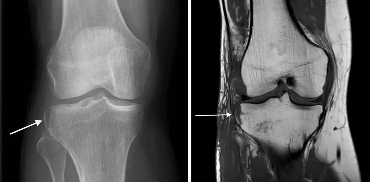
A 28-year-old man twisted his knee playing basketball. An AP radiograph of the knee and a coronal T1-weighted MRI image are shown. Which injury is most strongly associated with this finding?
- PCL tear
- Patellar dislocation
- Posterior root tear of the medial meniscus
- ACL tear
- Quadriceps tendon rupture
Show the answer
Answer: D. ACL tear
A small, vertically oriented flake of bone is lifted off the lateral tibial plateau just below the joint line, at the attachment of the lateral capsule. This is a Segond fracture, an avulsion by the anterolateral ligament and capsule, and it is very strongly associated with ACL tears and meniscal injuries, so MRI is recommended.
Key point: Segond fracture: vertical avulsion from the lateral tibial plateau just below the joint (anterolateral ligament and capsule) = ACL tear until proven otherwise; reverse Segond (medial) = PCL and medial meniscal injury; arcuate fragment of the fibular head = posterolateral corner.
Topic: Segond fracture. From Chapter 12: Knee: menisci, ligaments, extensor mechanism and cartilage. Practice the full block in RadQBoard.
9. Ultrasound behind the knee during a DVT scan
A 72-year-old man had a duplex ultrasound for calf swelling to look for deep vein thrombosis. The grayscale ultrasound image behind the knee shown here was obtained. What is the most likely diagnosis?
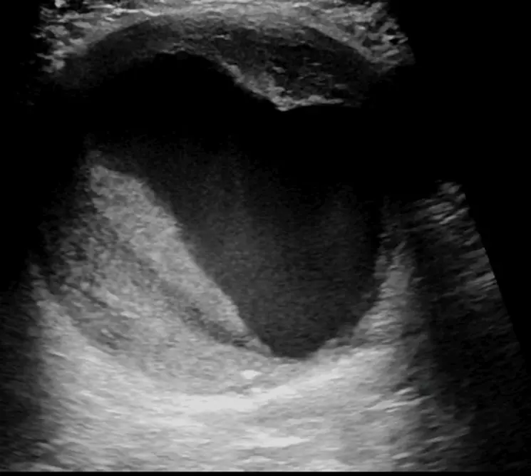
- Baker cyst
- Popliteal vein thrombosis
- Parameniscal cyst
- Soft-tissue sarcoma
- Popliteal artery aneurysm
Show the answer
Answer: E. Popliteal artery aneurysm
The popliteal artery is markedly widened, with echogenic mural thrombus lining its lumen. A dilated popliteal artery with layered thrombus is a popliteal artery aneurysm, which must be distinguished from a Baker cyst with Doppler.
Key point: Popliteal artery aneurysm: the most common peripheral arterial aneurysm, often bilateral and associated with abdominal aortic aneurysm; risks are thrombosis and distal embolization; Doppler distinguishes it from a Baker cyst.
Topic: Popliteal artery aneurysm. From Chapter 13: Knee osteochondral lesions and synovium, hip anatomy and hip fractures. Practice the full block in RadQBoard.
10. Shoulder MR arthrogram in an older man
A 72-year-old man has chronic shoulder pain and a soft swelling on top of his shoulder. Coronal fat-suppressed MR arthrogram images, obtained after injection of contrast into the glenohumeral joint, are shown. What do the findings indicate?
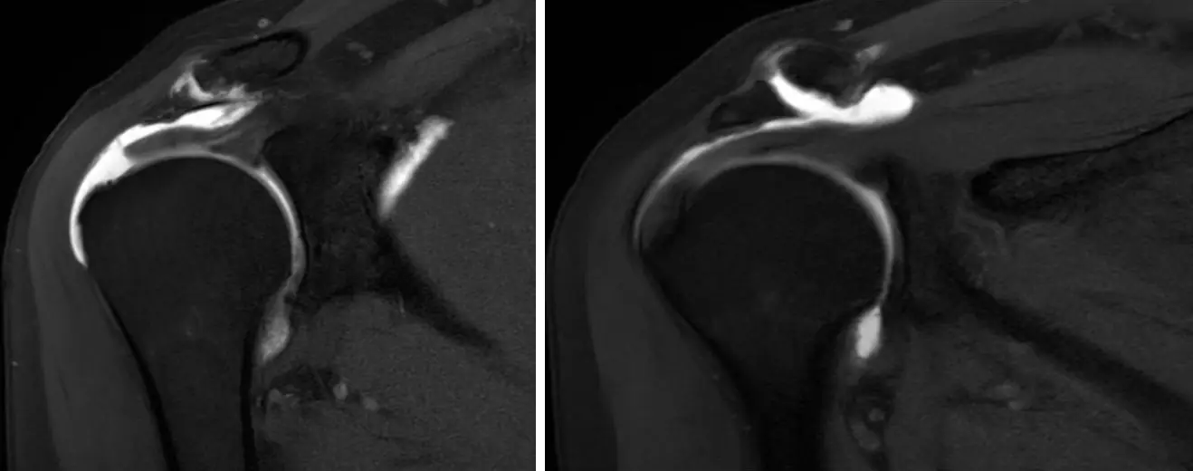
- Normal arthrogram with physiologic filling of the subacromial bursa and joint recesses
- Partial-thickness articular-surface cuff tear only
- Full-thickness cuff tear with fluid escaping through the AC joint (geyser sign)
- Septic arthritis of the AC joint
- Os acromiale
Show the answer
Answer: C. Full-thickness cuff tear with fluid escaping through the AC joint (geyser sign)
Contrast injected into the glenohumeral joint has passed through a full-thickness supraspinatus tear into the subacromial-subdeltoid bursa and continues up through the degenerated AC joint into a cyst above it. This geyser sign occurs with chronic, usually massive, rotator cuff tears and AC joint degeneration.
Key point: The subacromial-subdeltoid bursa does not normally communicate with the glenohumeral joint: arthrographic contrast in the bursa means a full-thickness cuff tear; extension through the AC joint into a superior cyst is the geyser sign of chronic massive tears.
Topic: Geyser sign. From Chapter 14: Hip fractures, osteonecrosis, impingement and the shoulder girdle. Practice the full block in RadQBoard.
Free with an account.