RadQBoard › Musculoskeletal › Chapter 10
Midfoot, hindfoot and ankle
Starting at the midfoot, this chapter covers Lisfranc and Chopart injuries, calcaneal and talar fractures, osteochondral lesions of the talus and tarsal coalition. It then turns to the ankle: the lateral and medial ligaments, the tendons around the ankle on MRI, and the syndesmosis with the fracture patterns it shapes.
Read all 10 pages and answer the chapter's 30 questions in the app. Free with an account.
What this chapter covers
Lisfranc injuryFree
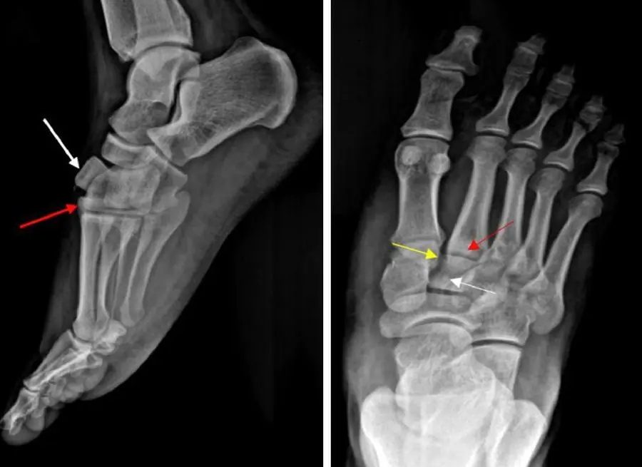
The second metatarsal base is the keystone of the midfoot: when it slips even a little from the middle cuneiform, the Lisfranc ligament may be torn.
Chopart injury and navicular osteonecrosis
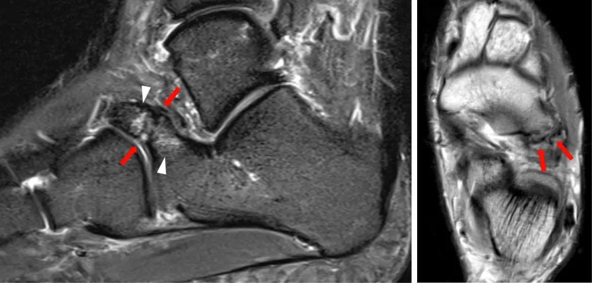
The Chopart joint is the talonavicular plus calcaneocuboid joint; the navicular can also die, in boys as Köhler disease and in women as Müller-Weiss disease.
Calcaneal fractures
A fall from a height: a flattened Böhler angle, a fractured posterior facet, and a lumbar spine to check.
Talar fractures
A talar neck fracture can cut off the blood to the body of the talus; a lucent band under the dome at 6–8 weeks (Hawkins sign) is good news.
Osteochondral lesion of the talus
A crescent under the talar dome; on MRI, fluid creeping under the fragment means it is unstable.
Tarsal coalition
An adolescent with a stiff, painful flatfoot: look for a C sign, a talar beak or an anteater nose.
Lateral ankle ligaments and sprains
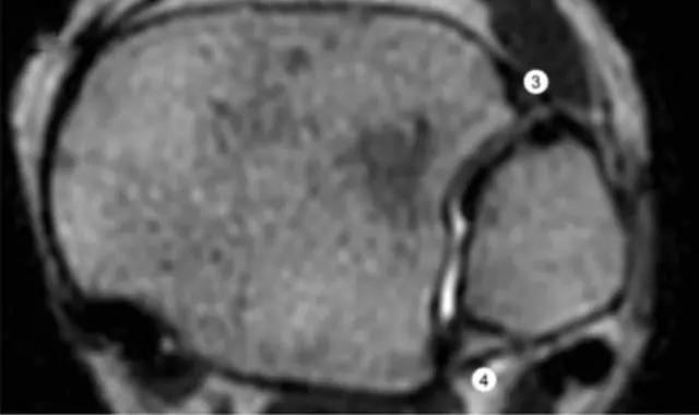
The ATFL tears first, the CFL next, and the PTFL almost never; a comma-shaped fibula tells you that you are at the level of the ATFL.
Ankle tendons on MRI
Front: Tom, Harry and Dick. Medial: Tom, Dick, ANd Harry. Lateral: the peroneals. Back: the Achilles.
Medial ankle: deltoid and spring ligaments
The deltoid and spring ligaments hold up the medial arch with the posterior tibial tendon; when the tendon fails, the foot flattens.
Syndesmosis and ankle fractures
The syndesmosis opens like a book from the front; the height of the fibular fracture (Weber A, B or C) tells you whether it is likely to be torn.
Further reading
- Siddiqui NA, Galizia MS, Almusa E, et al. Evaluation of the Tarsometatarsal Joint Using Conventional Radiography, CT, and MR Imaging. RadioGraphics. 2014;34(2):514-531. doi:10.1148/rg.342125215
- Walter WR, Hirschmann A, Alaia EF, et al. Normal Anatomy and Traumatic Injury of the Midtarsal (Chopart) Joint Complex: An Imaging Primer. RadioGraphics. 2019;39(1):136-152. doi:10.1148/rg.2019180102
- Smitaman E, Davis M. Hindfoot Fractures: Injury Patterns and Relevant Imaging Findings. RadioGraphics. 2022;42(3):661-682. doi:10.1148/rg.210167
- Newman JS, Newberg AH. Congenital Tarsal Coalition: Multimodality Evaluation with Emphasis on CT and MR Imaging. RadioGraphics. 2000;20(2):321-332. doi:10.1148/radiographics.20.2.g00mc03321
- Crim JR, Kjeldsberg KM. Radiographic Diagnosis of Tarsal Coalition. American Journal of Roentgenology. 2004;182(2):323-328. doi:10.2214/ajr.182.2.1820323
- Rosenberg ZS, Beltran J, Bencardino JT. MR Imaging of the Ankle and Foot. RadioGraphics. 2000;20(suppl_1):S153-S179. doi:10.1148/radiographics.20.suppl_1.g00oc26s153
- Taljanovic MS, Alcala JN, Gimber LH, et al. High-Resolution US and MR Imaging of Peroneal Tendon Injuries. RadioGraphics. 2015;35(1):179-199. doi:10.1148/rg.351130062
- Flores DV, Mejía Gómez C, Fernández Hernando M, et al. Adult Acquired Flatfoot Deformity: Anatomy, Biomechanics, Staging, and Imaging Findings. RadioGraphics. 2019;39(5):1437-1460. doi:10.1148/rg.2019190046
- da Silva LNM, Morimoto LR, Watanabe GG, et al. Distal Tibiofibular Syndesmosis: Anatomy, Biomechanics, Imaging Approach, and Postoperative Evaluation. RadioGraphics. 2026;46(2):e250065. doi:10.1148/rg.250065
Read all 10 pages and answer the chapter's 30 questions in the app. Free with an account.