RadQBoard

RadQBoard › Musculoskeletal › Chapter 10 › Lisfranc injury

Lisfranc injury

Musculoskeletal · Chapter 10: Midfoot, hindfoot and ankle · Page 1 of 10 · Free to read

The second metatarsal base is the keystone of the midfoot: when it slips even a little from the middle cuneiform, the Lisfranc ligament may be torn.

What it is

The Lisfranc joint is the tarsometatarsal joint. Its key stabilizer, the Lisfranc ligament, runs from the medial (first) cuneiform to the base of the second metatarsal. A Lisfranc injury ranges from a ligament sprain to a fracture-dislocation of the joint.

Patterns

Pattern Displacement
Homolateral All metatarsals shift laterally
Divergent The first metatarsal shifts medially, the second to fifth laterally
Isolated (partial) One or a few metatarsals displaced
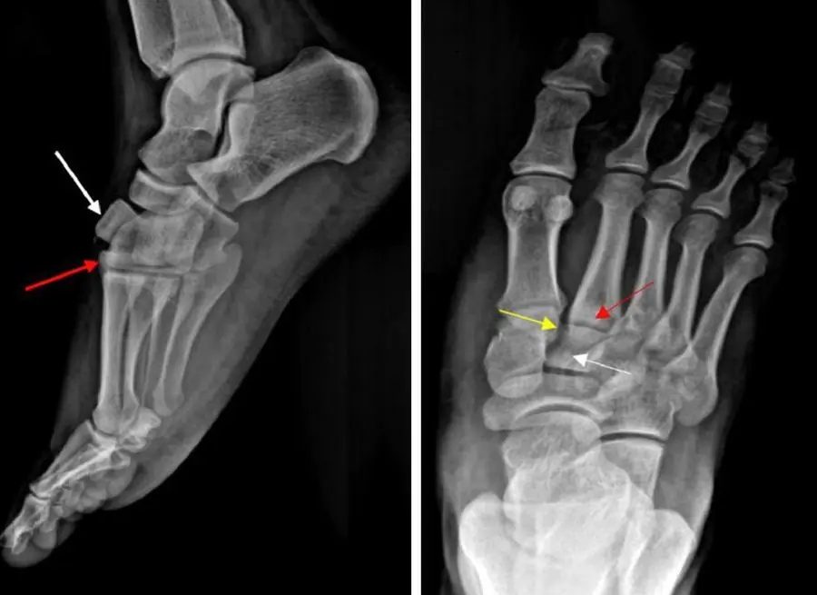
Lateral and frontal foot radiographs with white, red and yellow arrows at the midfoot
Lisfranc-type fracture-dislocation. Left, lateral radiograph: the intermediate cuneiform (white arrow) and the second metatarsal base (red arrow) are displaced dorsally, breaking the smooth dorsal outline of the tarsometatarsal joint. Right, frontal radiograph: the second metatarsal base is out of line (red arrow), with the displaced intermediate cuneiform (white arrow) and a fleck of bone between the medial cuneiform and the second metatarsal base (yellow arrow). Image: Gholipour M, Salimi M, Motamedi A, et al. A rare Lisfranc-type injury involving dorsal dislocation of the intermediate cuneiform and second metatarsal. Radiol Case Rep 2025;20:529-534 (Fig. 1A, C). CC BY 4.0. Changes: cropped; panel letters removed (2 of 3 panels); arrows from the original figure.

Radiographs

Check alignment on every foot radiograph, ideally weight-bearing, which is the most sensitive view:

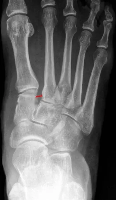
Weight-bearing frontal foot radiograph with a short red line between the medial cuneiform and the second metatarsal base
Subtle Lisfranc injury in a 21-year-old footballer, weight-bearing frontal (dorsoplantar) radiograph. The gap between the medial cuneiform and the base of the second metatarsal is widened (red line, more than 4 mm; normal is 2 mm or less), and the second metatarsal base no longer lines up with the middle cuneiform. Weight-bearing views unmask this kind of diastasis. Image: Crone AM, Omar IM, Bronson AM, et al. Guide to lower extremity radiologic measurements: part 3 ankle and foot. Skeletal Radiol 2026;55:1487-1511 (Fig. 19). CC BY 4.0. Changes: cropped; measurement label removed; measurement line from the original figure.
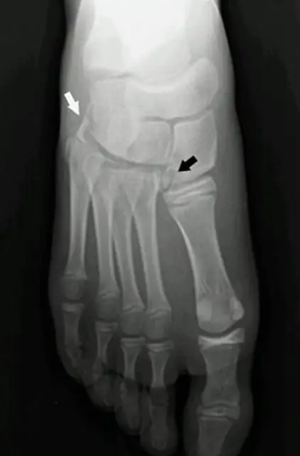
Frontal foot radiograph of a child with a black arrow between the first two metatarsal bases and a white arrow at the cuboid
Lisfranc fracture-dislocation in a child, frontal foot radiograph. Black arrow: the fleck sign, a small fragment avulsed from the lateral side of the medial cuneiform between the first and second metatarsal bases. White arrow: a cuboid fracture, with lateral subluxation of the second to fifth tarsometatarsal joints. Image: Ventura M, Ferreira A, Rodrigues D, et al. Pediatric Lisfranc fracture-dislocation: a case report. Cureus 2022;14:e29525 (Fig. 1). CC BY 4.0. Changes: cropped (1 of 3 panels); side marker and screen text removed; arrows from the original figure.

CT and MRI

CT shows fractures and subtle displacement; MRI shows the ligament itself when radiographs are normal but suspicion is high.

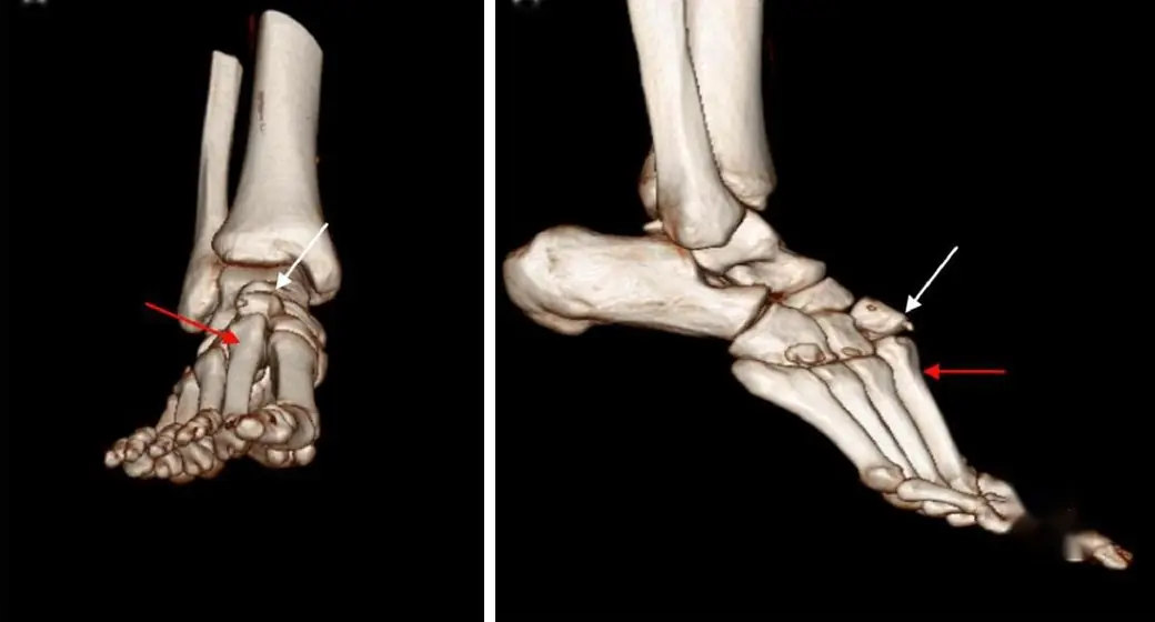
Two 3D CT reconstructions of the foot from the front and the side with white and red arrows at the midfoot
Same injury, 3D CT reconstructions from the front (left) and the side (right): the intermediate cuneiform (white arrows) and the second metatarsal base (red arrows) are dislocated dorsally. CT shows the full extent of fractures and displacement that radiographs underestimate. Image: Gholipour M, Salimi M, Motamedi A, et al. A rare Lisfranc-type injury involving dorsal dislocation of the intermediate cuneiform and second metatarsal. Radiol Case Rep 2025;20:529-534 (Fig. 2). CC BY 4.0. Changes: cropped; panel letters removed; viewer text removed; arrows from the original figure.

Further reading

  1. Siddiqui NA, Galizia MS, Almusa E, et al. Evaluation of the Tarsometatarsal Joint Using Conventional Radiography, CT, and MR Imaging. RadioGraphics. 2014;34(2):514-531. doi:10.1148/rg.342125215

Practice this in RadQBoard. Chapter 10 has 30 board-style questions with full explanations. Free with an account.
Open RadQBoard Chapter 10 overview

Written in original words by the RadQBoard team. Images are reused under open licenses, with the source, license and any changes under each one.