Musculoskeletal · Chapter 11: Ankle fractures, ankle tendons and the knee: trauma and menisci · Page 10 of 10 · Free to read
The handle flips into the notch to make a second PCL, or forward to make two anterior horns.
What it is
A bucket-handle tear is a displaced vertical longitudinal tear: the inner part of the meniscus (the handle) flips out of place, while the outer part (the bucket) stays attached. The medial meniscus is affected more often. The fragment can lock the knee.
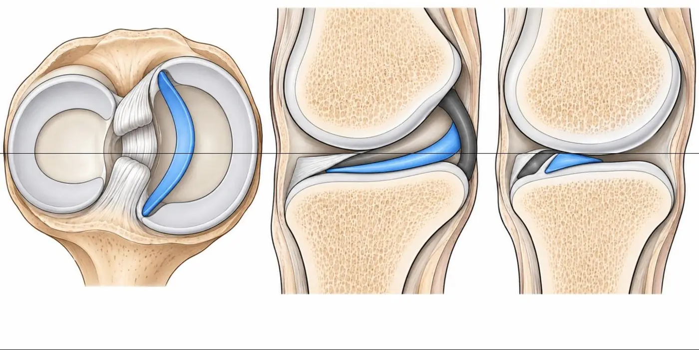
Illustration: a bucket-handle tear, displaced fragment in blue. Left, seen from above: the inner fragment (handle) of a vertical longitudinal tear of the medial meniscus has flipped into the intercondylar notch beside the cruciate ligaments, while the outer rim (bucket) stays attached. Middle, sagittal through the notch: the handle lies just below the PCL (dark band), the double PCL sign. Right, sagittal through the meniscus: when the handle flips forward it lies just behind the anterior horn, giving two triangles at the front and a small or absent posterior horn, the double delta sign. Illustration adapted from knee MRI (Nguyen JC, et al. Skeletal Radiol 2026, Fig. 16) (CC BY 4.0). CC BY 4.0. Changes: simplified drawing of the pattern in the source figure; no tracing. Source article.
MRI signs
Sign
Meaning
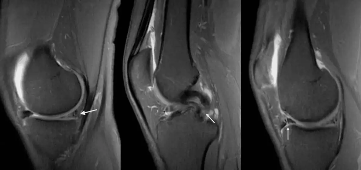
Absent bowtie
Fewer than two bowtie-shaped slices; the body is short because part of it is gone
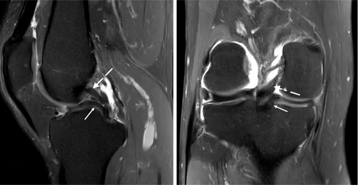
Double PCL
The handle lies in the intercondylar notch under and parallel to the PCL, like a second, smaller PCL
Double delta
The handle flips forward and lies just behind the native anterior horn, making two triangles
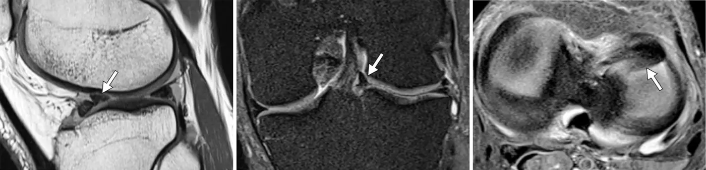
Fragment in the notch
On coronal images, a dark band in the intercondylar notch
Absent bow-tie sign: fewer than two bow-tie slices on sagittal images suggests that a fragment has displaced.
A medial posterior-horn fragment cannot flip all the way forward past an intact ACL, so it stays in the notch: double PCL suggests a medial tear with an intact ACL.
The lateral meniscus is not blocked by the ACL: double delta suggests a lateral tear (or a medial tear with a torn ACL).
Usually needs arthroscopic repair or partial meniscectomy, so the location and size of the displaced fragment should be reported.
Count the menisci: when a meniscus looks small, look for the missing piece.
Further reading
Nguyen JC, De Smet AA, Graf BK, et al. MR Imaging–based Diagnosis and Classification of Meniscal Tears. RadioGraphics. 2014;34(4):981-999. doi:10.1148/rg.344125202
Practice this in RadQBoard. Chapter 11 has 30 board-style questions with full explanations. Free with an account.