RadQBoard › Musculoskeletal › Chapter 12 › ACL tear: secondary signs, Segond fracture and reconstruction
ACL tear: secondary signs, Segond fracture and reconstruction
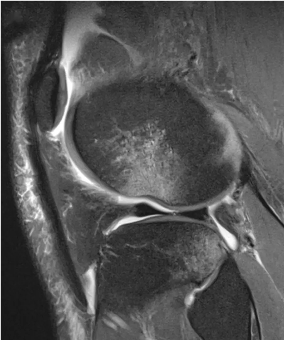
Kissing bruises on the lateral femoral condyle and posterior tibia, a buckled PCL and a sliver off the lateral tibia all point to a torn ACL.
What it is
When the ACL tears, the tibia slides forward and rotates, leaving indirect signs in the bones and other ligaments. These signs help when the ligament itself is hard to judge. After reconstruction, the graft position and scar tissue explain most problems.
Secondary signs on MRI
| Sign | Why |
|---|---|
| Pivot-shift bone contusions: lateral femoral condyle (middle, at the sulcus) and posterolateral tibial plateau | The tibia slides forward and the bones strike each other |
| Deep lateral femoral notch (more than about 1.5 mm) | Impaction at the condylopatellar sulcus |
| Anterior tibial translation (more than about 5–7 mm) | Loss of the ACL restraint |
| Buckled PCL | The tibia moves forward and the PCL loses its tension |
| Uncovered posterior horn of the lateral meniscus | Forward shift of the tibia |
- Deep lateral femoral notch: a sulcus deeper than about 1.5–2 mm on the lateral radiograph suggests an ACL tear.
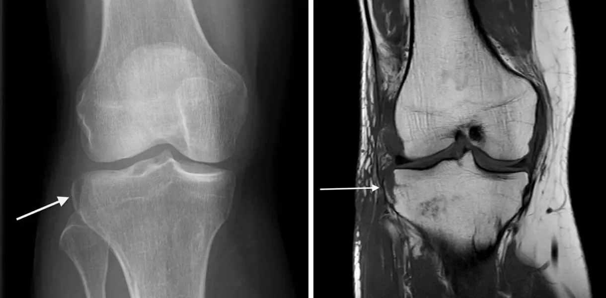
Segond fracture
- A small, vertically oriented avulsion from the lateral tibial plateau, just below the joint line, from the lateral capsule (anterolateral ligament) and iliotibial band fibers.
- Easy to miss on a radiograph, but very strongly linked to ACL tear and meniscal injury: recommend MRI.
- Separate it from a Gerdy tubercle avulsion (iliotibial band, more anterior) and an arcuate fracture of the fibular head (posterolateral corner).
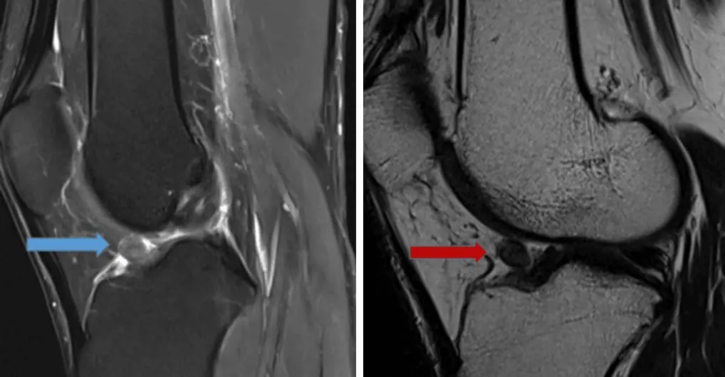
ACL reconstruction
- The graft should run parallel to and behind Blumensaat's line.
- Too vertical: unstable knee. Too anterior on the tibia: the graft hits the notch roof in extension (impingement).
- Cyclops lesion: a nodule of fibrous scar in front of the graft in Hoffa's fat pad that blocks full extension.
- Other problems: graft tear, tunnel widening and ganglia, hardware failure.
Further reading
- Gentili A, Seeger LL, Yao L, et al. Anterior cruciate ligament tear: indirect signs at MR imaging. Radiology. 1994;193(3):835-840. doi:10.1148/radiology.193.3.7972834
- Hayes CW, Brigido MK, Jamadar DA, et al. Mechanism-based Pattern Approach to Classification of Complex Injuries of the Knee Depicted at MR Imaging. RadioGraphics. 2000;20(suppl_1):S121-S134. doi:10.1148/radiographics.20.suppl_1.g00oc21s121
Practice this in RadQBoard. Chapter 12 has 30 board-style questions with full explanations. Free with an account.
Written in original words by the RadQBoard team. Images are reused under open licenses, with the source, license and any changes under each one.