RadQBoard › Musculoskeletal › Chapter 12
Knee: menisci, ligaments, extensor mechanism and cartilage
This chapter completes the menisci with radial tears, parameniscal cysts and the discoid meniscus, then covers each knee ligament: the ACL with its secondary signs and reconstruction, the PCL, the MCL and the posterolateral corner. It ends with the extensor mechanism, lateral patellar dislocation, Osgood-Schlatter disease and how to describe cartilage defects.
Free page: ACL tear: secondary signs, Segond fracture and reconstruction
Read all 10 pages and answer the chapter's 30 questions in the app. Free with an account.
What this chapter covers
Radial tears and parameniscal cysts
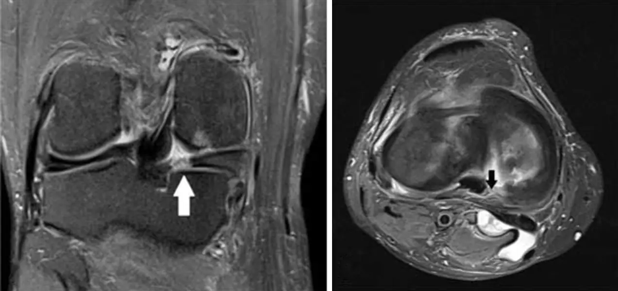
A radial tear cuts across the meniscus from its free edge; on sagittal images the cleft marches, or the horn vanishes as a ghost.
Discoid meniscus
Too much meniscus: a bowtie that persists for three or more sagittal slices, usually laterally, in a child with a clicking knee.
Anterior cruciate ligament
A normal ACL is a taut, striated band parallel to the intercondylar roof; when it tears, look for the bone bruises it leaves behind.
ACL tear: secondary signs, Segond fracture and reconstructionFree
Kissing bruises on the lateral femoral condyle and posterior tibia, a buckled PCL and a sliver off the lateral tibia all point to a torn ACL.
Posterior cruciate ligament
The PCL is thick, dark and rarely torn; when it is, it usually stretches and swells rather than snapping.
Medial collateral ligament
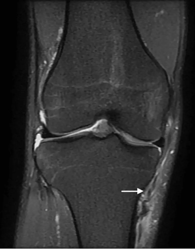
Valgus force pulls the MCL apart from sprain to complete tear; years later, a fleck of calcium by the medial femoral condyle may be all that is left.
Lateral knee: LCL, posterolateral corner and iliotibial band
From back to front at the lateral knee: biceps femoris, LCL, iliotibial band. A torn posterolateral corner is a near-emergency.
Extensor mechanism: quadriceps and patellar tendons
Quadriceps tear: patella sits low. Patellar tendon tear: patella rides high. Both: the patient cannot straighten the knee.
Lateral patellar dislocation and Osgood-Schlatter disease
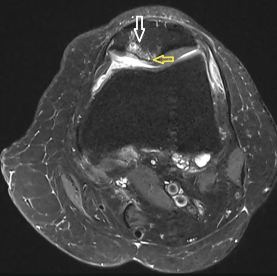
Even after it pops back, a dislocated patella leaves kissing bruises on the medial patella and lateral femoral condyle and a torn medial retinaculum.
Knee cartilage
Describe every cartilage defect by where, how deep, and how big; full-thickness defects and delamination change management.
Further reading
- Nguyen JC, De Smet AA, Graf BK, et al. MR Imaging–based Diagnosis and Classification of Meniscal Tears. RadioGraphics. 2014;34(4):981-999. doi:10.1148/rg.344125202
- Gentili A, Seeger LL, Yao L, et al. Anterior cruciate ligament tear: indirect signs at MR imaging. Radiology. 1994;193(3):835-840. doi:10.1148/radiology.193.3.7972834
- Hayes CW, Brigido MK, Jamadar DA, et al. Mechanism-based Pattern Approach to Classification of Complex Injuries of the Knee Depicted at MR Imaging. RadioGraphics. 2000;20(suppl_1):S121-S134. doi:10.1148/radiographics.20.suppl_1.g00oc21s121
- Lundquist RB, Matcuk GR Jr, Schein AJ, et al. Posteromedial Corner of the Knee: The Neglected Corner. RadioGraphics. 2015;35(4):1123-1137. doi:10.1148/rg.2015140166
- Vinson EN, Major NM, Helms CA. The Posterolateral Corner of the Knee. American Journal of Roentgenology. 2008;190(2):449-458. doi:10.2214/ajr.07.2051
- Flores DV, Mejía Gómez C, Pathria MN. Layered Approach to the Anterior Knee: Normal Anatomy and Disorders Associated with Anterior Knee Pain. RadioGraphics. 2018;38(7):2069-2101. doi:10.1148/rg.2018180048
- Watts RE, Gorbachova T, Fritz RC, et al. Patellar Tracking: An Old Problem with New Insights. RadioGraphics. 2023;43(6):e220177. doi:10.1148/rg.220177
- Pathria MN, Chung CB, Resnick DL. Acute and Stress-related Injuries of Bone and Cartilage: Pertinent Anatomy, Basic Biomechanics, and Imaging Perspective. Radiology. 2016;280(1):21-38. doi:10.1148/radiol.16142305
Read all 10 pages and answer the chapter's 30 questions in the app. Free with an account.