RadQBoard › Musculoskeletal › Chapter 13
Knee osteochondral lesions and synovium, hip anatomy and hip fractures
The knee section covers osteochondral lesions, synovial disease such as tenosynovial giant cell tumor (PVNS), and the Baker cyst with its mimics. The hip section starts with radiographic landmarks and the blood supply of the femoral head, then covers acetabular fractures, dislocation, the occult fracture after a fall in an older adult, and femoral neck fractures.
Read all 10 pages and answer the chapter's 30 questions in the app. Free with an account.
What this chapter covers
Osteochondral lesions of the knee
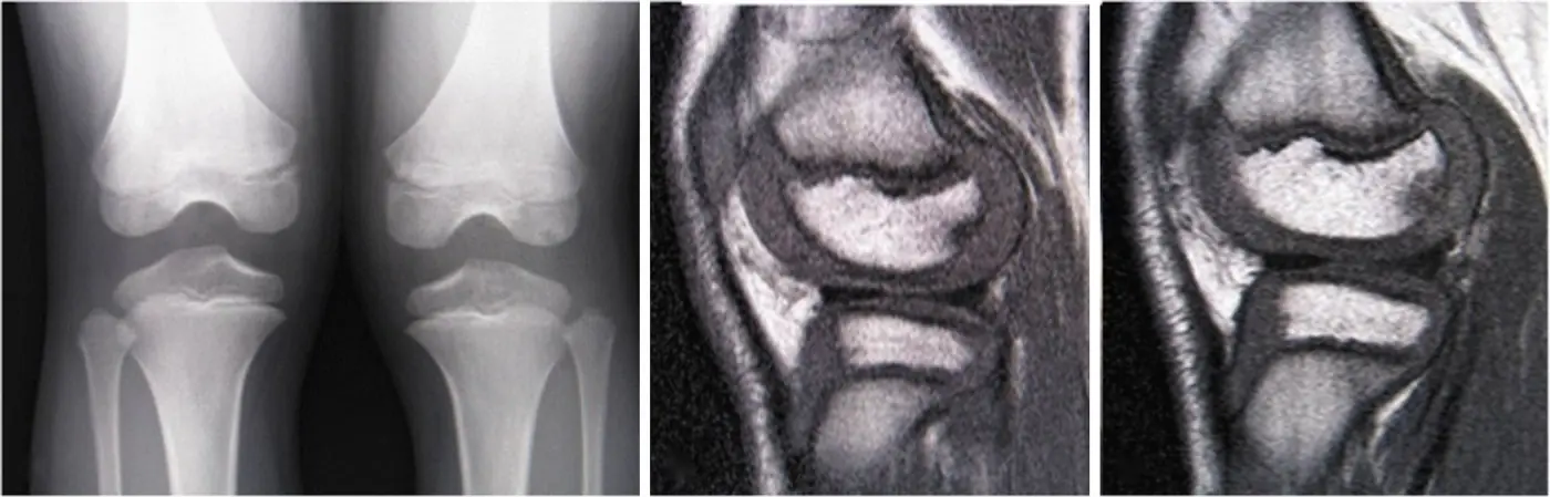
One umbrella term, four causes: OCD in teenagers, fracture after trauma, insufficiency fracture in older adults, and osteonecrosis. Fluid under the fragment means unstable.
Tenosynovial giant cell tumor (PVNS) and lipoma arborescens
Dark on every sequence and blooming on gradient echo: hemosiderin-laden synovium. Fat-signal fronds: lipoma arborescens.
Baker cyst and tennis leg
A Baker cyst comes out between the semimembranosus and the medial gastrocnemius (M&M); a pulsating popliteal mass may be an aneurysm instead.
Hip anatomy: acetabulum, labrum and radiographic landmarks
The iliopectineal line traces the anterior column and the ilioischial line the posterior column; know the trochanters and who attaches where.
Acetabular fractures
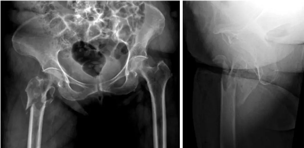
Break in the iliopectineal line = anterior column; break in the ilioischial line = posterior column; the posterior wall is the piece most often broken.
Proximal femur: anatomy and blood supply
The femoral head lives on the medial femoral circumflex artery; fractures inside the capsule threaten it, fractures outside do not.
Hip dislocation and femoral head fractures
About 9 in 10 dislocations are posterior: the femur is flexed, adducted and internally rotated, and the head sits up and back.
Imaging an elderly fall: occult hip fracture
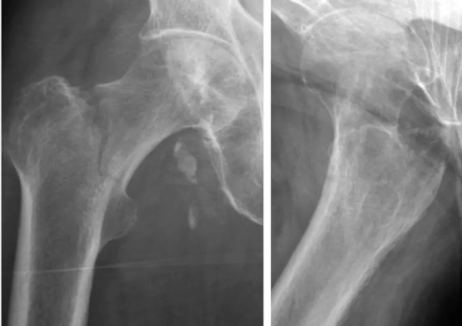
Up to 1 in 10 hip fractures is invisible on the first radiographs; MRI finds essentially all of them.
Femoral neck fractures
Valgus impacted counts as nondisplaced and gets screws; varus displaced gets a new hip.
Basicervical and greater trochanter fractures
Basicervical fractures sit just outside the capsule and do well with a sliding screw; a "tiny" greater trochanter fracture is often the tip of an intertrochanteric iceberg.
Further reading
- Gorbachova T, Melenevsky Y, Cohen M, et al. Osteochondral Lesions of the Knee: Differentiating the Most Common Entities at MRI. RadioGraphics. 2018;38(5):1478-1495. doi:10.1148/rg.2018180044
- Murphey MD, Rhee JH, Lewis RB, et al. Pigmented Villonodular Synovitis: Radiologic-Pathologic Correlation. RadioGraphics. 2008;28(5):1493-1518. doi:10.1148/rg.285085134
- Flores DV, Mejía Gómez C, Estrada-Castrillón M, et al. MR Imaging of Muscle Trauma: Anatomy, Biomechanics, Pathophysiology, and Imaging Appearance. RadioGraphics. 2018;38(1):124-148. doi:10.1148/rg.2018170072
- Scheinfeld MH, Dym AA, Spektor M, et al. Acetabular Fractures: What Radiologists Should Know and How 3D CT Can Aid Classification. RadioGraphics. 2015;35(2):555-577. doi:10.1148/rg.352140098
- Sheehan SE, Shyu JY, Weaver MJ, et al. Proximal Femoral Fractures: What the Orthopedic Surgeon Wants to Know. RadioGraphics. 2015;35(5):1563-1584. doi:10.1148/rg.2015140301
- Haj-Mirzaian A, Eng J, Khorasani R, et al. Use of Advanced Imaging for Radiographically Occult Hip Fracture in Elderly Patients: A Systematic Review and Meta-Analysis. Radiology. 2020;296(3):521-531. doi:10.1148/radiol.2020192167
- Oka M, Monu JUV. Prevalence and Patterns of Occult Hip Fractures and Mimics Revealed by MRI. American Journal of Roentgenology. 2004;182(2):283-288. doi:10.2214/ajr.182.2.1820283
Read all 10 pages and answer the chapter's 30 questions in the app. Free with an account.