RadQBoard › Musculoskeletal › Chapter 14
Hip fractures, osteonecrosis, impingement and the shoulder girdle
Hip fractures continue with intertrochanteric, subtrochanteric, stress and atypical femoral fractures, followed by femoral head osteonecrosis and transient bone marrow edema. The chapter then covers femoroacetabular impingement and labral tears before moving to the shoulder: AC separation, the bursae and impingement, and rotator cuff anatomy.
Free page: Femoral head osteonecrosis (avascular necrosis)
Read all 10 pages and answer the chapter's 30 questions in the app. Free with an account.
What this chapter covers
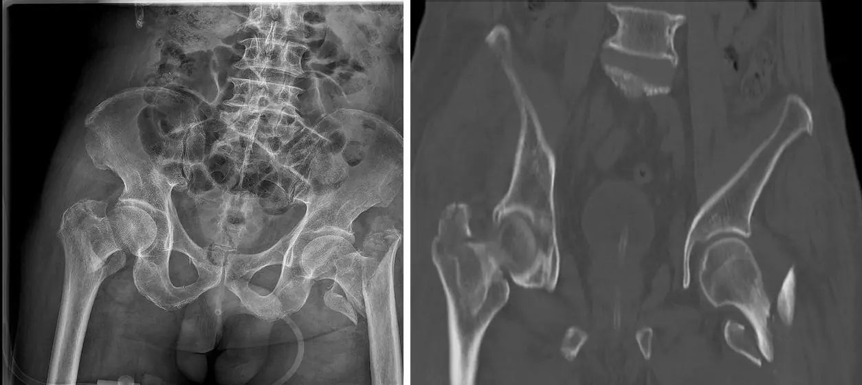
Intertrochanteric and subtrochanteric fractures
A clean two-part intertrochanteric fracture gets a sliding screw; comminution or a broken lateral wall gets a nail.
Femoral neck stress fracture
In a runner with groin pain: the compression side (medial) heals with rest; the tension side (lateral) can displace and needs aggressive care.
Atypical femoral fracture
A small beak of thickened lateral cortex below the lesser trochanter can be the only warning before the femur snaps transversely.
Femoral head osteonecrosis (avascular necrosis)Free
A serpiginous dark rim on T1, a double line on T2, in both hips of a patient on steroids; find it before the head collapses.
Transient bone marrow edema of the hip
Severe hip pain, osteopenic femoral head, diffuse edema on MRI, and nothing else: a diagnosis made only after looking hard for a fracture or osteonecrosis.
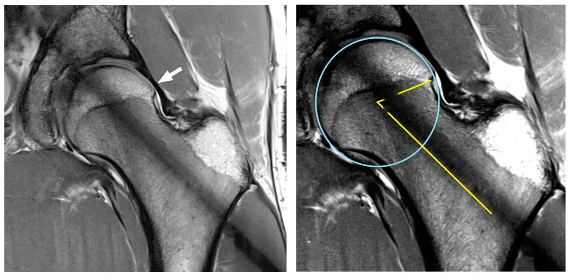
Femoroacetabular impingement
Cam is a bump on the femoral head-neck junction; pincer is too much acetabular cover. Shapes are common; impingement is a clinical diagnosis.
Acetabular labral tears
Usually anterosuperior and usually from impingement; MR arthrography shows contrast cutting under or into the labrum.
Shoulder bones and acromioclavicular separation
Grade the AC injury by the AC gap and the coracoclavicular distance; above about 25% widening of the CC space, the CC ligaments are torn.
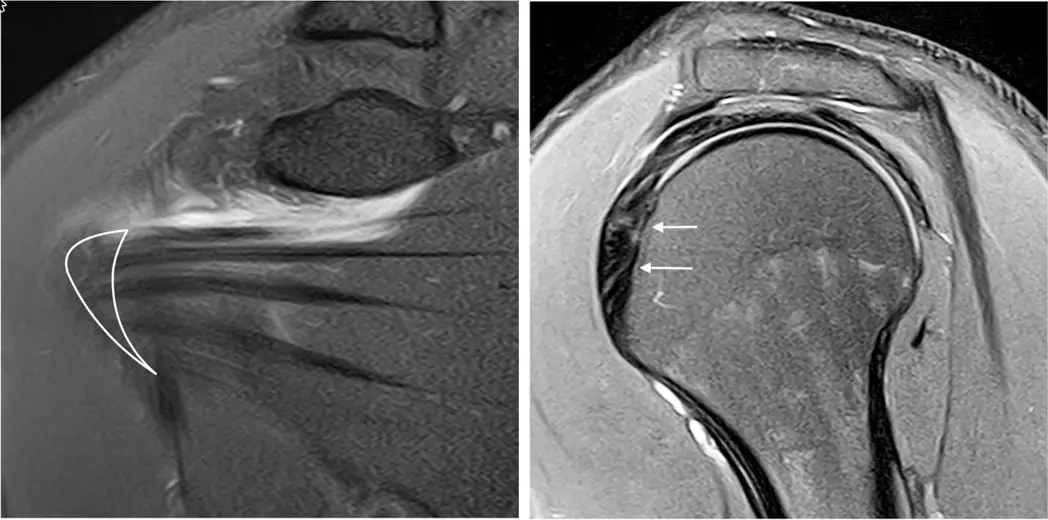
Shoulder bursae and impingement
Fluid from the joint into the subacromial-subdeltoid bursa means a full-thickness cuff tear; a hooked acromion or os acromiale may narrow the outlet.
Rotator cuff anatomy
Three cuff tendons go to the greater tuberosity from behind the scapula; one, the subscapularis, goes to the lesser tuberosity from the front.
Further reading
- Sheehan SE, Shyu JY, Weaver MJ, et al. Proximal Femoral Fractures: What the Orthopedic Surgeon Wants to Know. RadioGraphics. 2015;35(5):1563-1584. doi:10.1148/rg.2015140301
- Hegazi TM, Belair JA, McCarthy EJ, et al. Sports Injuries about the Hip: What the Radiologist Should Know. RadioGraphics. 2016;36(6):1717-1745. doi:10.1148/rg.2016160012
- La Rocca Vieira R, Rosenberg ZS, Allison MB, et al. Frequency of Incomplete Atypical Femoral Fractures in Asymptomatic Patients on Long-Term Bisphosphonate Therapy. American Journal of Roentgenology. 2012;198(5):1144-1151. doi:10.2214/ajr.11.7442
- Murphey MD, Foreman KL, Klassen-Fischer MK, et al. From the Radiologic Pathology Archives Imaging of Osteonecrosis: Radiologic-Pathologic Correlation. RadioGraphics. 2014;34(4):1003-1028. doi:10.1148/rg.344140019
- Laborie LB, Lehmann TG, Engesæter IØ, et al. Prevalence of Radiographic Findings Thought to Be Associated with Femoroacetabular Impingement in a Population-based Cohort of 2081 Healthy Young Adults. Radiology. 2011;260(2):494-502. doi:10.1148/radiol.11102354
- Flores DV, Foster RCB, Sampaio ML, et al. Hip Capsulolabral Complex: Anatomy, Disease, MRI Features, and Postoperative Appearance. RadioGraphics. 2024;44(2):e230144. doi:10.1148/rg.230144
- Flores DV, Goes PK, Gómez CM, et al. Imaging of the Acromioclavicular Joint: Anatomy, Function, Pathologic Features, and Treatment. RadioGraphics. 2020;40(5):1355-1382. doi:10.1148/rg.2020200039
- Lee MH, Sheehan SE, Orwin JF, et al. Comprehensive Shoulder US Examination: A Standardized Approach with Multimodality Correlation for Common Shoulder Disease. RadioGraphics. 2016;36(6):1606-1627. doi:10.1148/rg.2016160030
Read all 10 pages and answer the chapter's 30 questions in the app. Free with an account.