RadQBoard › Musculoskeletal › Chapter 14 › Femoral head osteonecrosis (avascular necrosis)
Femoral head osteonecrosis (avascular necrosis)
A serpiginous dark rim on T1, a double line on T2, in both hips of a patient on steroids; find it before the head collapses.
What it is
Death of the subchondral bone and marrow of the femoral head from interrupted blood supply. The dead bone can collapse, leading to secondary osteoarthritis.
Causes
- Trauma: displaced femoral neck fractures, hip dislocation, even transient subluxation in athletes.
- Systemic (often bilateral): corticosteroids, alcohol, sickle cell disease, Gaucher disease, transplant, radiation, pancreatitis, lupus.
- Clues: a small calcified spleen suggests sickle cell disease; splenomegaly suggests Gaucher disease.
Radiographs
- Normal early.
- Later: patchy sclerosis and lucency, then a subchondral lucent crescent (collapse beginning), flattening of the head, and finally secondary arthritis.
- A crescent sign (a subchondral lucent line) means a subchondral fracture; collapse is starting or about to.
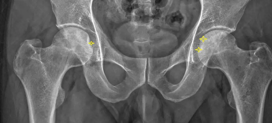
MRI (most sensitive)
- A geographic subchondral area in the anterosuperior femoral head outlined by a serpentine low-signal rim on T1.
- Double-line sign on T2: a dark outer line and a bright inner line; considered characteristic.
- Marrow edema in about half, linked with pain and early collapse.
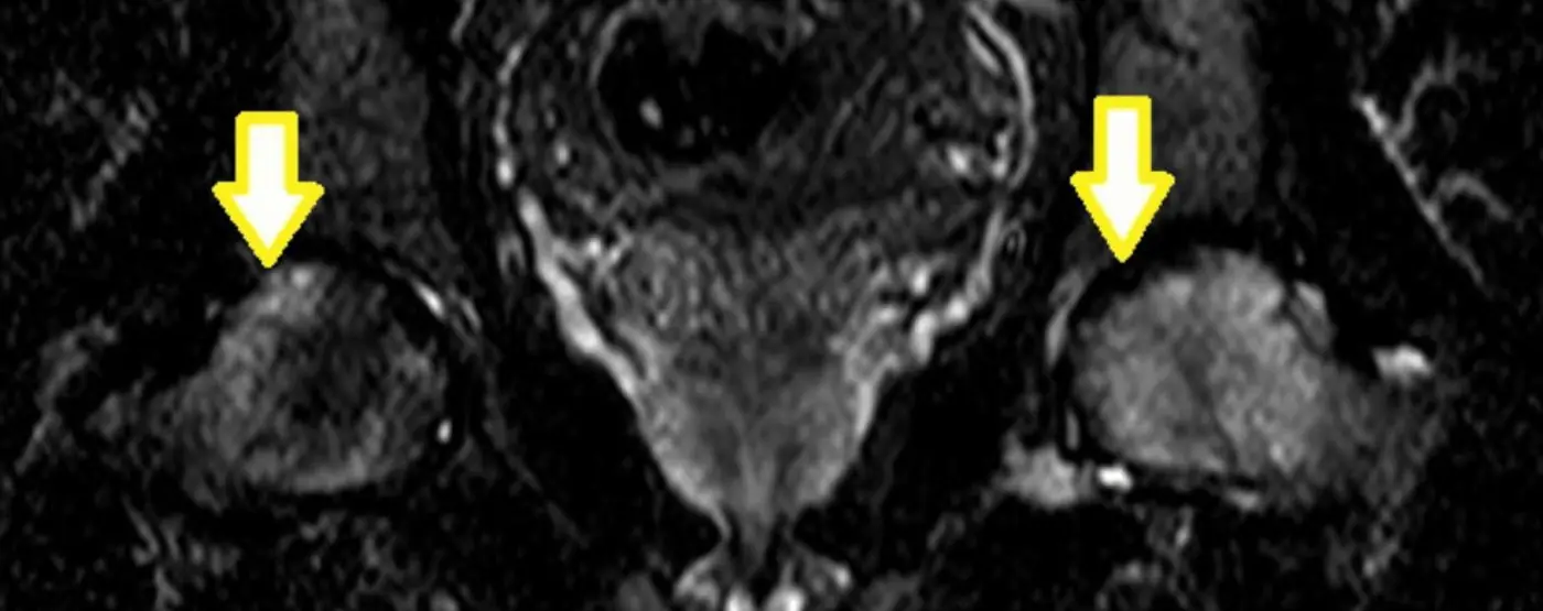
What to report
- Size of the necrotic area (fraction of the weight-bearing surface), whether the subchondral bone plate has collapsed, and secondary arthritis. Examine the other hip.
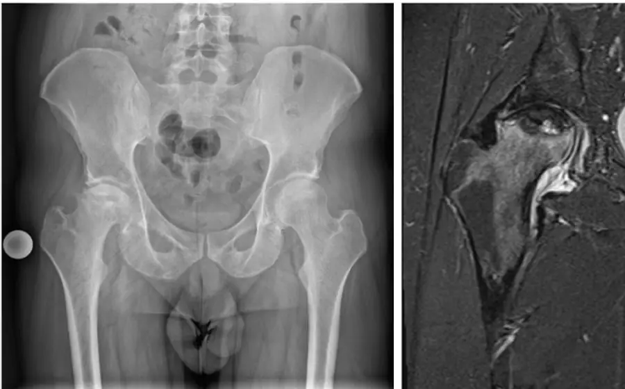
Treatment
- Before collapse: protected weight-bearing, medications, core decompression.
- After collapse with arthritis: arthroplasty.
Further reading
- Murphey MD, Foreman KL, Klassen-Fischer MK, et al. From the Radiologic Pathology Archives Imaging of Osteonecrosis: Radiologic-Pathologic Correlation. RadioGraphics. 2014;34(4):1003-1028. doi:10.1148/rg.344140019
Practice this in RadQBoard. Chapter 14 has 30 board-style questions with full explanations. Free with an account.
Written in original words by the RadQBoard team. Images are reused under open licenses, with the source, license and any changes under each one.