RadQBoard › Musculoskeletal › Chapter 15
Shoulder: rotator cuff, capsule, biceps, labrum and dislocation
The chapter opens with the three planes of shoulder MRI, then follows rotator cuff disease from tendinosis and partial tears to full-thickness tears with retraction and fatty atrophy. It covers the glenohumeral ligaments, the rotator interval and frozen shoulder, the long head of the biceps, and the normal labral variants that mimic tears. It closes with dislocation in each direction, the Hill-Sachs and Bankart lesions, and the named labroligamentous injuries.
Read all 10 pages in the app. Free with an account.
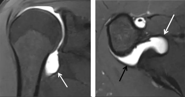
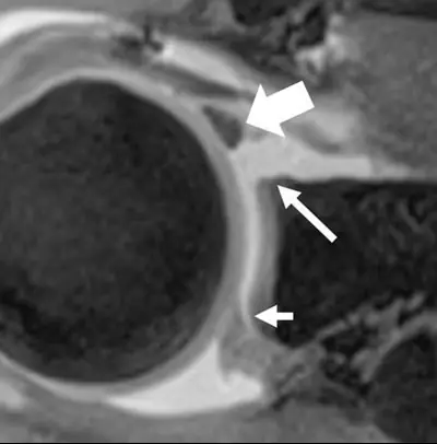
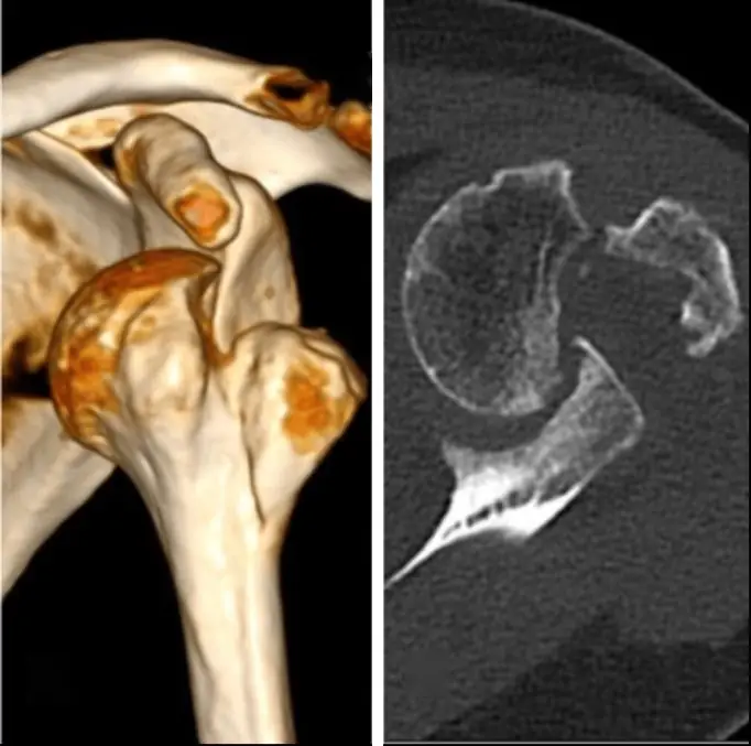
What this chapter covers
Normal shoulder MRI anatomy
Three planes, three questions: oblique coronal for the supraspinatus, axial for the labrum and biceps, oblique sagittal for the muscles and the arch.
Rotator cuff tendinosis and partial-thickness tears
Most partial tears start on the articular side of the supraspinatus at its footprint: the rim-rent or PASTA lesion.
Full-thickness rotator cuff tears and muscle atrophy
A gap from the joint to the bursa, a retracted tendon end, and fat in the muscle: what the surgeon needs to know before repair.
Shoulder ligaments, rotator interval and adhesive capsulitis
The inferior glenohumeral ligament holds the shoulder in abduction and external rotation; a thick, stiff capsule and a filled-in rotator interval mean frozen shoulder.
Long head of the biceps: tears and dislocation
An empty bicipital groove means the tendon has torn and retracted or has slipped medially, usually through a torn subscapularis.
Shoulder instability and the glenoid labrum
A shallow socket made stable by soft tissue; 95% of instability is anterior, and some labral gaps are normal variants.
Anterior shoulder dislocation: Hill-Sachs and Bankart
When the head pops forward and down, the back of the humeral head hits the front of the glenoid: a Hill-Sachs dent on the humerus and a Bankart injury on the glenoid.
Anterior labroligamentous injuries
Many names for one idea: where the anteroinferior labrum and the inferior glenohumeral ligament tore, and whether the periosteum held.
Posterior and inferior shoulder dislocation
Posterior dislocation after a seizure is easy to miss on the frontal view: look for the lightbulb, the overlap on the Grashey view and the trough line.
Shoulder dislocation: putting it together
A one-page comparison of the three directions of dislocation and the injuries each leaves behind.
Further reading
- de Jesus JO, Parker L, Frangos AJ, et al. Accuracy of MRI, MR Arthrography, and Ultrasound in the Diagnosis of Rotator Cuff Tears: A Meta-Analysis. American Journal of Roentgenology. 2009;192(6):1701-1707. doi:10.2214/ajr.08.1241
- Petchprapa CN, Beltrán L, Jazrawi LM, et al. The Rotator Interval: A Review of Anatomy, Function, and Normal and Abnormal MRI Appearance. American Journal of Roentgenology. 2010;195(3):567-576. doi:10.2214/ajr.10.4406
- Gondim Teixeira PA, Balaj C, Chanson A, et al. Adhesive Capsulitis of the Shoulder: Value of Inferior Glenohumeral Ligament Signal Changes on T2-Weighted Fat-Saturated Images. American Journal of Roentgenology. 2012;198(6):W589-W596. doi:10.2214/ajr.11.7453
- Roy E, Cheyne I, Andrews GT, et al. Beyond the Cuff: MR Imaging of Labroligamentous Injuries in the Athletic Shoulder. Radiology. 2016;278(2):316-332. doi:10.1148/radiol.2015150364
- Sharifi A, Siebert MJ, Chhabra A. How to Measure Glenoid Bone Stock and Version and Why It Is Important: A Practical Guide. RadioGraphics. 2020;40(6):1671-1683. doi:10.1148/rg.2020200008
- Beltran J, Rosenberg ZS, Chandnani VP, et al. Glenohumeral instability: evaluation with MR arthrography. RadioGraphics. 1997;17(3):657-673. doi:10.1148/radiographics.17.3.9153704
- Saupe N, White LM, Bleakney RR, et al. Acute Traumatic Posterior Shoulder Dislocation: MR Findings. Radiology. 2008;248(1):185-193. doi:10.1148/radiol.2481071003
Read all 10 pages in the app. Free with an account.