RadQBoard › Musculoskeletal › Chapter 16
Shoulder instability lesions, nerve entrapment, elbow and forearm trauma
It starts with how to find instability lesions on MRI (the Hill-Sachs notch, the GLOM sign and humeral avulsion of the inferior glenohumeral ligament), then covers posterior instability, the thrower's Bennett lesion, SLAP tears, GLAD and paralabral cysts. Nerve problems around the shoulder follow, from suprascapular and axillary nerve entrapment to brachial neuritis. The second half covers elbow dislocation and the terrible triad, the fat pad sign and radial head fracture, the forearm fracture-dislocations and distal radius fractures.
Read all 10 pages in the app. Free with an account.
What this chapter covers
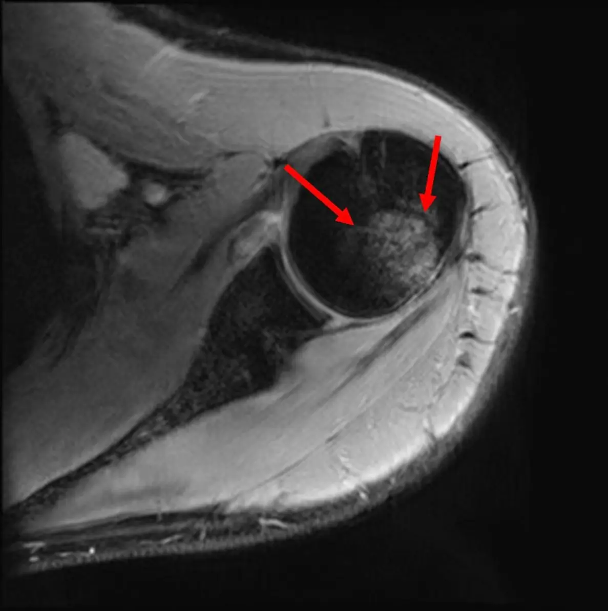
Finding Hill-Sachs, GLOM and HAGL on MRI
Scroll the top three axial slices for a notch in the back of the humeral head; a dark ball in the joint may be a torn labrum that has rolled up.
Posterior instability and the Bennett lesion
Less than 1 in 20 unstable shoulders, but the mirror images of anterior lesions appear: reverse Bankart, reverse Hill-Sachs and posterior HAGL.
SLAP tears
A tear of the superior labrum running front to back across the biceps anchor; on coronal images, a bright line that curves laterally is a tear, a smooth medial one is a recess.
GLAD lesion and paralabral cysts
A labral tear with a chip of cartilage, and a cyst next to the labrum that says "there is a tear here" even when you cannot see it.
Suprascapular nerve entrapment
Compression at the suprascapular notch weakens both the supraspinatus and infraspinatus; at the spinoglenoid notch, only the infraspinatus.
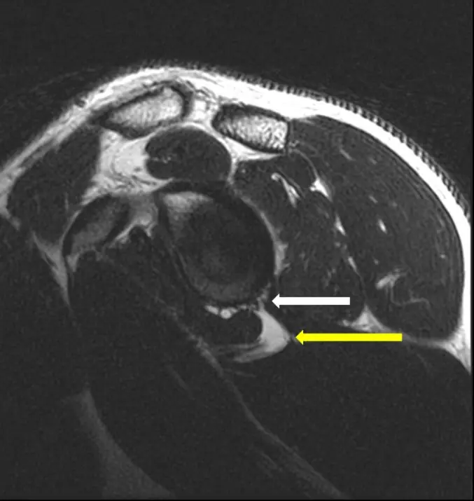
Quadrilateral space syndrome and Parsonage-Turner syndrome
Teres minor atrophy alone: the axillary nerve in the quadrilateral space. Patchy cuff and deltoid edema without a cause: brachial neuritis.
Elbow dislocation and the terrible triad
The elbow is the second most dislocated joint; dislocation plus a radial head fracture plus a coronoid fracture is the terrible triad, and the coronoid fragment may be tiny.
Radial head fracture and the fat pad sign
A visible posterior fat pad on a true lateral view means an effusion and, after trauma, a fracture until proven otherwise.
Forearm fracture-dislocations: Monteggia, Galeazzi, Essex-Lopresti
The forearm is a ring: if one bone breaks and shortens, a joint must dislocate. Always image the elbow and the wrist.
Distal radius fractures
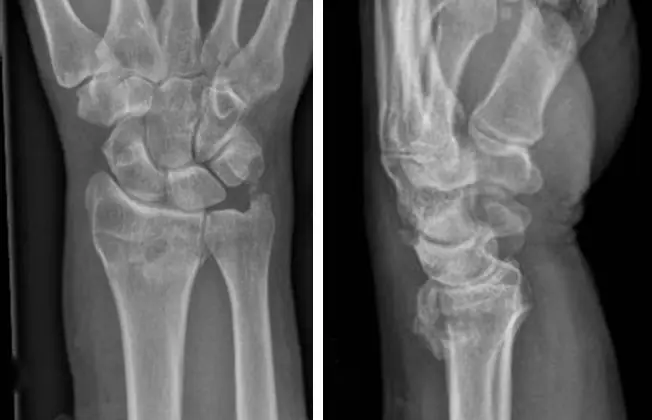
Colles tilts back, Smith tilts forward, Barton shears off a rim with the carpus, Hutchinson splits off the styloid.
Further reading
- Roy EA, Cheyne I, Andrews GT, et al. Beyond the Cuff: MR Imaging of Labroligamentous Injuries in the Athletic Shoulder. Radiology. 2016;278(2):316-332. doi:10.1148/radiol.2015150364
- Saupe N, White LM, Bleakney R, et al. Acute Traumatic Posterior Shoulder Dislocation: MR Findings. Radiology. 2008;248(1):185-193. doi:10.1148/radiol.2481071003
- Symanski JS, Subhas N, Babb J, et al. Diagnosis of Superior Labrum Anterior-to-Posterior Tears by Using MR Imaging and MR Arthrography: A Systematic Review and Meta-Analysis. Radiology. 2017;285(1):101-113. doi:10.1148/radiol.2017162681
- Jin W, Ryu KN, Kwon SH, et al. MR Arthrography in the Differential Diagnosis of Type II Superior Labral Anteroposterior Lesion and Sublabral Recess. American Journal of Roentgenology. 2006;187(4):887-893. doi:10.2214/ajr.05.0955
- Scalf RE, Wenger DE, Frick MA, et al. MRI Findings of 26 Patients with Parsonage-Turner Syndrome. American Journal of Roentgenology. 2007;189(1):W39-W44. doi:10.2214/ajr.06.1136
- Sheehan SE, Dyer GS, Sodickson AD, et al. Traumatic Elbow Injuries: What the Orthopedic Surgeon Wants to Know. RadioGraphics. 2013;33(3):869-888. doi:10.1148/rg.333125176
- Major NM, Crawford ST. Elbow Effusions in Trauma in Adults and Children: Is There an Occult Fracture?. American Journal of Roentgenology. 2002;178(2):413-418. doi:10.2214/ajr.178.2.1780413
- Goldfarb CA, Yin Y, Gilula LA, et al. Wrist Fractures: What the Clinician Wants to Know. Radiology. 2001;219(1):11-28. doi:10.1148/radiology.219.1.r01ap1311
Read all 10 pages in the app. Free with an account.