RadQBoard › Musculoskeletal › Chapter 17
Elbow tendons and ligaments, wrist and hand
At the elbow, this chapter covers the ulnar collateral ligament in throwers and tears of the triceps and distal biceps tendons. The wrist and hand follow: a map of named injuries, carpal alignment and instability patterns, perilunate and lunate dislocation, scaphoid fracture and its blood supply, triquetral fracture and Kienböck disease. It ends with the extensor compartments, the triangular fibrocartilage complex and finger tendon injuries.
Read all 10 pages in the app. Free with an account.
What this chapter covers
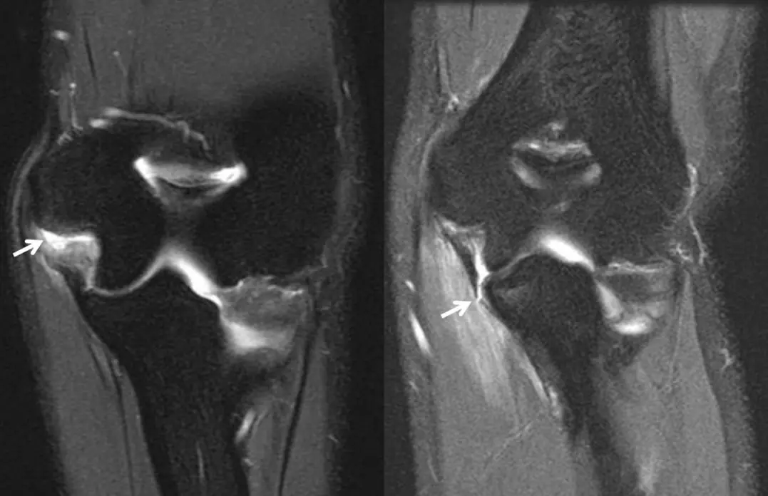
Elbow ulnar collateral ligament tear
Pitchers stress the inner elbow with every throw; the ligament peels off the sublime tubercle and contrast slips under it as a sideways T.
Triceps and distal biceps tendon tears
A fleck of bone above the olecranon after a fall on the hand is a triceps avulsion; a retracted biceps tendon means the lacertus fibrosus is torn too.
Hand and wrist injuries: a map
Each named hand injury lives in a predictable place; knowing the map tells you where to look.
Carpal alignment: arcs of Gilula, DISI and VISI
Three smooth arcs on the PA view, and a straight line of radius, lunate and capitate on the lateral view; a tilted lunate means a torn ligament.
Perilunate and lunate dislocation, scapholunate ligament
In perilunate dislocation the lunate stays with the radius and the capitate leaves; in lunate dislocation the capitate stays and the lunate tips out like a spilled teacup.
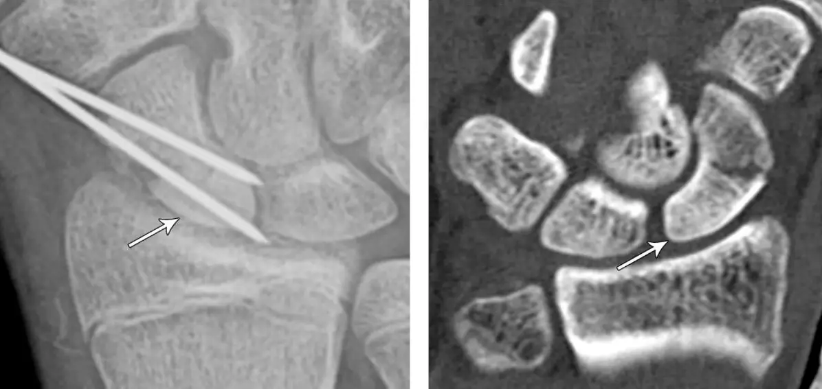
Scaphoid fracture
The most commonly broken carpal bone, often invisible at first; the blood flows from distal to proximal, so the proximal pole dies.
Triquetral fracture and Kienböck disease
A dorsal chip on the lateral wrist is the triquetrum; a small, dense, collapsed lunate with a short ulna is Kienböck.
Wrist extensor compartments and de Quervain tenosynovitis
Six dorsal compartments from radial to ulnar; fluid and thickening around the first, in a new mother, is de Quervain.
Triangular fibrocartilage complex
The disc between ulna and carpus: traumatic tears are slits and avulsions; degenerative tears thin the disc and wear the lunate.
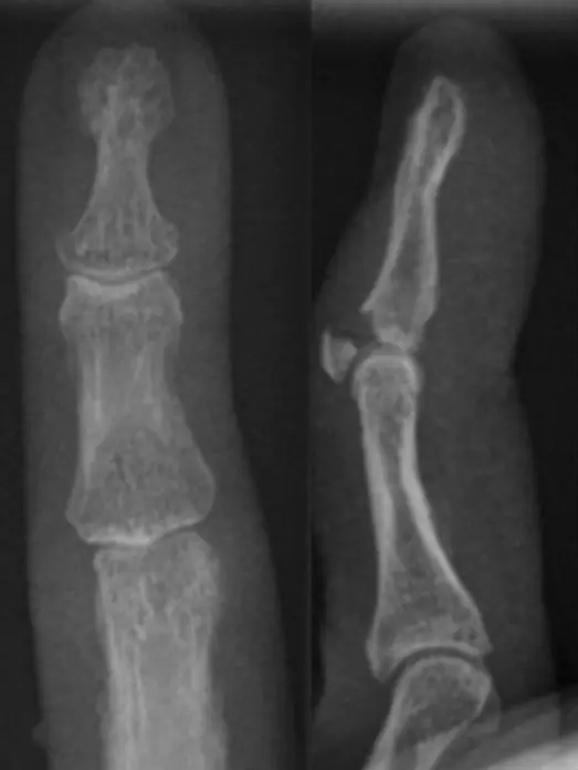
Finger tendon injuries: mallet, jersey and boutonniere
A drooping fingertip is mallet; a fingertip that won't bend is jersey; a bent PIP with an overextended DIP is boutonniere.
Further reading
- Goes PK, Flores DV, Damer A, et al. Shoulder and Elbow Injuries in Adult Overhead Throwers: Imaging Review. RadioGraphics. 2023;43(12):e230094. doi:10.1148/rg.230094
- Fitzgerald SW, Curry DR, Erickson SJ, et al. Distal biceps tendon injury: MR imaging diagnosis. Radiology. 1994;191(1):203-206. doi:10.1148/radiology.191.1.8134571
- Cockenpot E, Lefebvre G, Demondion X, et al. Imaging of Sports-related Hand and Wrist Injuries: Sports Imaging Series. Radiology. 2016;279(3):674-692. doi:10.1148/radiol.2016150995
- Weintraub MD, Hansford BG, Stilwill SE, et al. Avulsion Injuries of the Hand and Wrist. RadioGraphics. 2020;40(1):163-180. doi:10.1148/rg.2020190085
- Theumann NH, Etechami G, Duvoisin B, et al. Association between Extrinsic and Intrinsic Carpal Ligament Injuries at MR Arthrography and Carpal Instability at Radiography: Initial Observations. Radiology. 2006;238(3):950-957. doi:10.1148/radiol.2383050013
- Goldfarb CA, Yin Y, Gilula LA, et al. Wrist Fractures: What the Clinician Wants to Know. Radiology. 2001;219(1):11-28. doi:10.1148/radiology.219.1.r01ap1311
- Memarsadeghi M, Breitenseher MJ, Schaefer-Prokop C, et al. Occult Scaphoid Fractures: Comparison of Multidetector CT and MR Imaging—Initial Experience. Radiology. 2006;240(1):169-176. doi:10.1148/radiol.2401050412
- Oneson SR, Scales LM, Timins ME, et al. MR imaging interpretation of the Palmer classification of triangular fibrocartilage complex lesions. RadioGraphics. 1996;16(1):97-106. doi:10.1148/radiographics.16.1.97
- Burns JE, Tanaka T, Ueno T, et al. Pitfalls That May Mimic Injuries of the Triangular Fibrocartilage and Proximal Intrinsic Wrist Ligaments at MR Imaging. RadioGraphics. 2011;31(1):63-78. doi:10.1148/rg.311105114
- Clavero JA, Alomar X, Monill JM, et al. MR Imaging of Ligament and Tendon Injuries of the Fingers. RadioGraphics. 2002;22(2):237-256. doi:10.1148/radiographics.22.2.g02mr11237
Read all 10 pages in the app. Free with an account.