RadQBoard › Musculoskeletal › Chapter 18
Thumb and hand fractures, arthrography and musculoskeletal biopsy
The first part finishes the hand: the thumb ulnar collateral ligament and the Stener lesion, and the Bennett, Rolando, boxer's and volar plate fractures. The rest turns to procedures: how diagnostic and therapeutic joint injections are done safely with a low fluoroscopy dose, what a contrast leak means in the shoulder and wrist, and how to plan bone, spine and soft-tissue biopsies with the surgeon.
Read all 6 pages in the app. Free with an account.
What this chapter covers
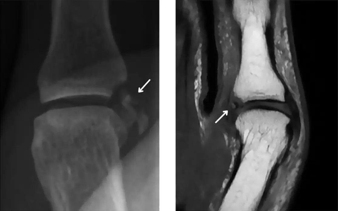
Gamekeeper's thumb and the Stener lesion
A torn thumb UCL heals in a cast, unless the adductor aponeurosis gets caught under it: the yo-yo on a string that needs surgery.
Bennett, Rolando, boxer's and volar plate fractures
Two thumb base fractures (one piece or many), a fifth metacarpal neck from punching, and a tiny volar chip at the PIP joint from hyperextension.
Arthrography: principles, safety and dose
Dilute gadolinium for MR arthrograms, a time-out before every needle, and fluoroscopy kept as short, small and low as possible.
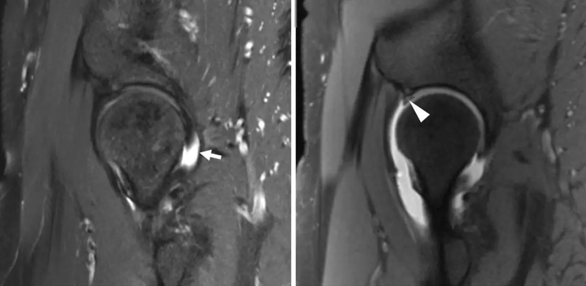
Shoulder, hip and wrist arthrograms
How much to inject and what a leak means: shoulder into the bursa = cuff tear; wrist into the DRUJ = TFC tear, into the midcarpal joint = intercarpal ligament tear.
Musculoskeletal biopsy: planning and soft-tissue biopsy
Image fully first, plan the route with the orthopedic oncologist, never cross an uninvolved compartment, and target the part that enhances.
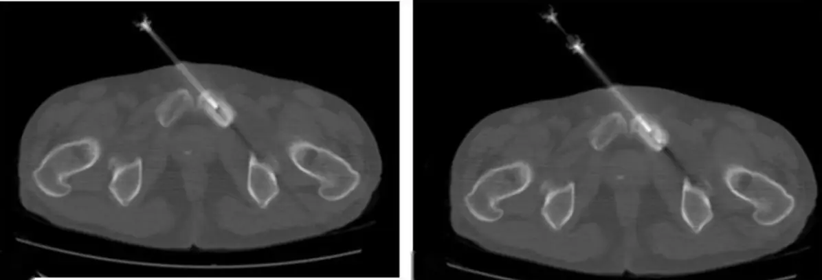
Bone, vertebral and osteomyelitis biopsy
Most bone biopsies are CT-guided; go through the pedicle in the spine; lytic lesions give more tissue than sclerotic ones; biopsy confirms the bug, not the diagnosis, in osteomyelitis.
Further reading
- Ebrahim FS, De Maeseneer M, Jager T, et al. US Diagnosis of UCL Tears of the Thumb and Stener Lesions: Technique, Pattern-based Approach, and Differential Diagnosis. RadioGraphics. 2006;26(4):1007-1020. doi:10.1148/rg.264055117
- Weintraub MD, Hansford BG, Stilwill SE, et al. Avulsion Injuries of the Hand and Wrist. RadioGraphics. 2020;40(1):163-180. doi:10.1148/rg.2020190085
- Steinbach LS, Palmer WE, Schweitzer ME. Special Focus Session: MR Arthrography. RadioGraphics. 2002;22(5):1223-1246. doi:10.1148/radiographics.22.5.g02se301223
- Kamel SI, Rosas HG, Gorbachova T. Local and Systemic Side Effects of Corticosteroid Injections for Musculoskeletal Indications. American Journal of Roentgenology. 2024;222(3):e2330458. doi:10.2214/ajr.23.30458
- Meek RD, Mills MK, Hanrahan CJ, et al. Pearls and Pitfalls for Soft-Tissue and Bone Biopsies: A Cross-Institutional Review. RadioGraphics. 2020;40(1):266-290. doi:10.1148/rg.2020190089
- Anderson MW, Temple HT, Dussault RG, et al. Compartmental anatomy: relevance to staging and biopsy of musculoskeletal tumors. American Journal of Roentgenology. 1999;173(6):1663-1671. doi:10.2214/ajr.173.6.10584817
- Debs P, Boutin RD, Smith SE, et al. Chronic Nonspinal Osteomyelitis in Adults: Consensus Recommendations on Percutaneous Bone Biopsies from the Society of Academic Bone Radiologists. Radiology. 2024;311(1):e231348. doi:10.1148/radiol.231348
Read all 6 pages in the app. Free with an account.