RadQBoard › Musculoskeletal › Chapter 11
Ankle fractures, ankle tendons and the knee: trauma and menisci
Ankle fractures are explained through the common injury sequence and the named fractures, followed by Achilles, posterior tibial and anterior tibial tendon injuries and the deltoid ligament. At the knee, the chapter covers dislocation, patellar and tibial plateau fractures, normal meniscal anatomy, and horizontal, vertical and bucket-handle tears.
Free page: Bucket-handle tears
Read all 10 pages and answer the chapter's 30 questions in the app. Free with an account.
What this chapter covers
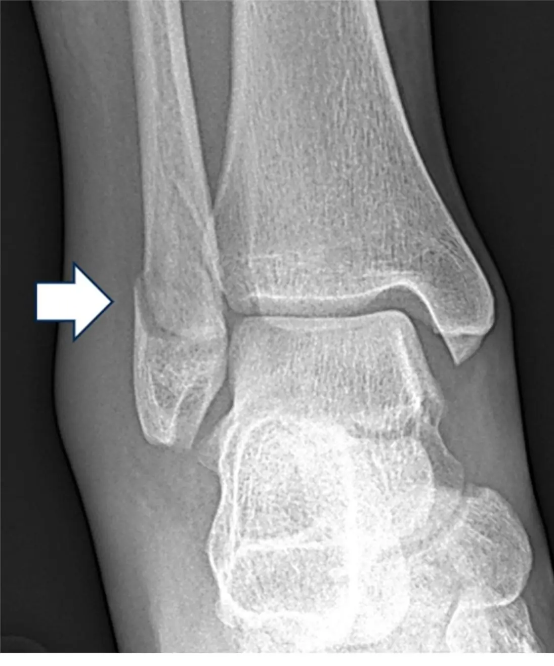
Supination-external rotation ankle fractures
Nine of ten ankle fractures follow the same four steps around the ankle, so the injury you can see predicts the ligaments you cannot.
Named ankle fractures
Tillaux and triplane in teenagers, Wagstaffe-Le Fort at the fibula, Maisonneuve up at the knee, and the pilon crushing the plafond.
Achilles tendon injury
The most commonly injured ankle tendon: a lost tendon outline and a cloudy Kager fat pad on the lateral radiograph.
Posterior and anterior tibial tendon injuries
A failing posterior tibial tendon flattens the arch in older adults; a ruptured anterior tibial tendon presents as a lump in front of the ankle.
Deltoid ligament injury
The deep deltoid is a single striated band from tibia to talus; a sliver of bone off the medial talus may be the only radiographic clue that it is torn.
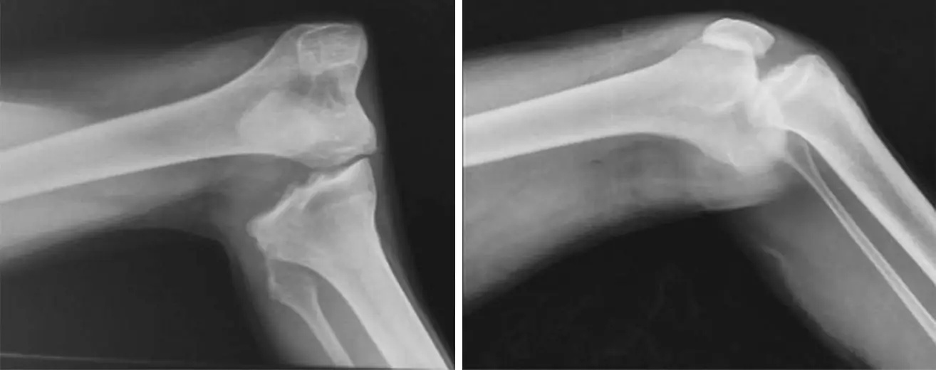
Knee dislocation, patellar and tibial plateau fractures
A dislocated knee threatens the popliteal artery even with normal pulses; a patella in two pieces may be a fracture or a bipartite variant.
Meniscal anatomy and normal MRI
Two C-shaped cushions, a red outer zone that can heal, and two meniscofemoral ligaments that pretend to be tears.
Myxoid degeneration and horizontal tears
Signal inside the meniscus only counts as a tear if it reaches the surface.
Vertical longitudinal tears
A vertical tear runs along the curve of the meniscus at a constant distance from its edge, from top surface to bottom surface.
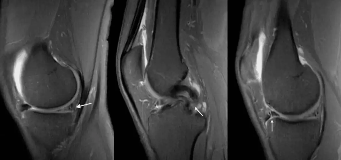
Bucket-handle tearsFree
The handle flips into the notch to make a second PCL, or forward to make two anterior horns.
Further reading
- Okanobo H, Khurana B, Sheehan S, et al. Simplified Diagnostic Algorithm for Lauge-Hansen Classification of Ankle Injuries. RadioGraphics. 2012;32(2):E71-E84. doi:10.1148/rg.322115017
- Schweitzer ME, Karasick D. MR Imaging of Disorders of the Achilles Tendon. American Journal of Roentgenology. 2000;175(3):613-625. doi:10.2214/ajr.175.3.1750613
- Schweitzer ME, Karasick D. MR Imaging of Disorders of the Posterior Tibialis Tendon. American Journal of Roentgenology. 2000;175(3):627-635. doi:10.2214/ajr.175.3.1750627
- Flores DV, Mejía Gómez C, Fernández Hernando M, et al. Adult Acquired Flatfoot Deformity: Anatomy, Biomechanics, Staging, and Imaging Findings. RadioGraphics. 2019;39(5):1437-1460. doi:10.1148/rg.2019190046
- Markhardt BK, Gross JM, Monu J. Schatzker Classification of Tibial Plateau Fractures: Use of CT and MR Imaging Improves Assessment. RadioGraphics. 2009;29(2):585-597. doi:10.1148/rg.292085078
- Nguyen JC, De Smet AA, Graf BK, et al. MR Imaging–based Diagnosis and Classification of Meniscal Tears. RadioGraphics. 2014;34(4):981-999. doi:10.1148/rg.344125202
Read all 10 pages and answer the chapter's 30 questions in the app. Free with an account.