RadQBoard › Musculoskeletal › Chapter 8 › Paget disease
Paget disease
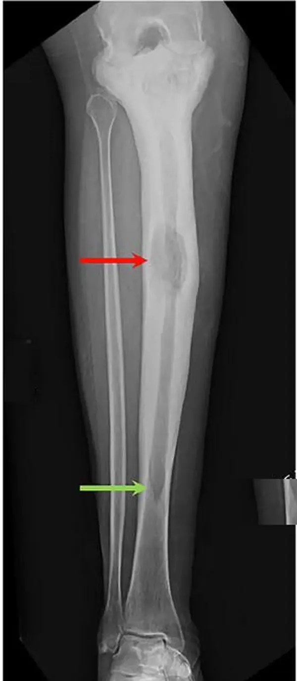
A caricature of normal bone in an older adult: bigger, thicker cortex, coarse trabeculae, and a lytic front shaped like a blade of grass.
What it is
Paget disease is a disorder of bone remodeling in older adults: overactive osteoclasts resorb bone, and disorganized new bone replaces it. The cause is unknown; a viral (paramyxovirus) trigger has been proposed.
- Symptoms: pain, enlargement or bowing of bones, compression of cranial nerves (deafness), and rarely high-output heart failure.
- Alkaline phosphatase is high; calcium and phosphate are normal.
Where it strikes
The skull, spine and pelvis most often, then the ends of the femur, proximal tibia and proximal humerus; ribs and scapula less often.
Three phases (they overlap)
| Phase | Picture |
|---|---|
| I. Lytic | Active resorption: lucent areas |
| II. Mixed | Resorption and new bone together |
| III. Sclerotic | Dense, thickened bone |
Imaging by site
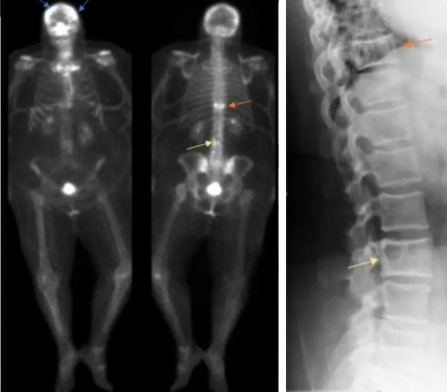
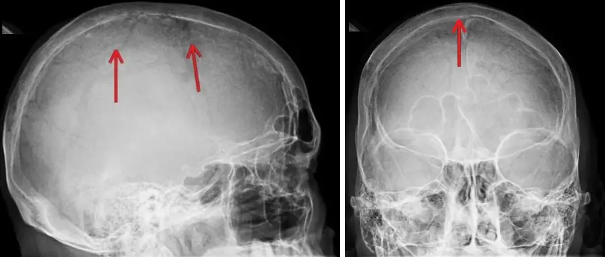
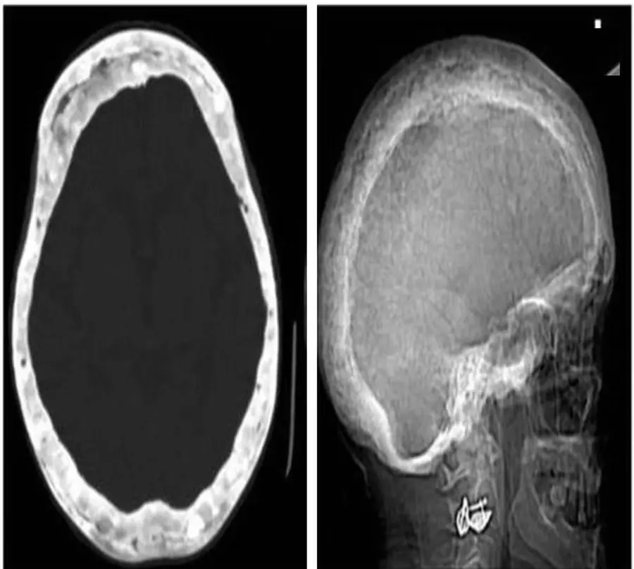
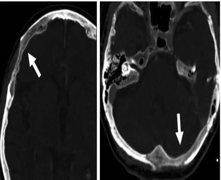
- Skull: lytic phase osteoporosis circumscripta, a sharply marginated lucent area; mixed phase cotton-wool skull, patchy cloud-like sclerosis with a thickened calvarium.
- Spine: picture-frame vertebra (dense thick rim around the whole body, which is also enlarged) or an ivory vertebra (uniformly dense).
- Pelvis: asymmetric coarse trabeculae and thickened iliopectineal and ilioischial lines; acetabular protrusio can follow.
- Long bones: the lytic phase starts at the end of the bone and advances along the shaft with a sharp, V-shaped blade-of-grass (flame-shaped) front, which separates it from a tumor. Later: bowing and coxa vara.
- Bone scan: intense uptake that outlines the whole involved bone.
- A bone scan is very hot in active disease and shows every site at once. On MRI, preserved fatty marrow argues against sarcoma.
Complications
- Fractures (including transverse fractures on the convex side of a bowed bone), osteoarthritis, basilar invagination, spinal stenosis.
- Sarcoma (rare; osteosarcoma most often), suggested by new pain, a lytic destructive area and a soft-tissue mass.
Further reading
- Smith SE, Murphey MD, Motamedi K, et al. From the Archives of the AFIP: Radiologic Spectrum of Paget Disease of Bone and Its Complications with Pathologic Correlation. RadioGraphics. 2002;22(5):1191-1216. doi:10.1148/radiographics.22.5.g02se281191
Practice this in RadQBoard. Chapter 8 has 30 board-style questions with full explanations. Free with an account.
Written in original words by the RadQBoard team. Images are reused under open licenses, with the source, license and any changes under each one.