RadQBoard

RadQBoard › Musculoskeletal › Chapter 7 › Contiguous osteomyelitis and the diabetic foot

Contiguous osteomyelitis and the diabetic foot

Musculoskeletal · Chapter 7: Nerve and other soft-tissue tumors, joint and bone infection · Page 9 of 10 · Free to read

An ulcer over a bone with a track down to it is osteomyelitis; a swollen midfoot without an ulcer is usually Charcot joint.

What it is

In adults, osteomyelitis most often spreads from a nearby soft-tissue infection, breaking through the periosteum and cortex before reaching the marrow. The classic setting is a diabetic foot ulcer: neuropathy leads to unnoticed trauma and ulcers, and poor blood supply slows healing.

Where it strikes

Pressure points under ulcers: the calcaneus, the first and fifth metatarsal heads, and the distal phalanx of the great toe.
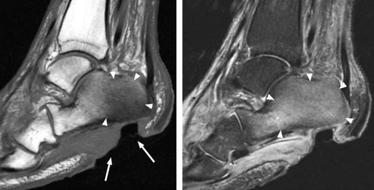
Sagittal T1 and STIR MRI of the heel with arrows at a plantar ulcer and arrowheads around the calcaneus
Calcaneal osteomyelitis under a heel ulcer, 48-year-old woman with diabetes. Sagittal T1 (left): a large plantar ulcer (arrows) reaches the inferior calcaneus, whose fatty marrow is replaced by confluent low signal (arrowheads). Sagittal STIR (right): matching marrow edema (arrowheads). Image: Alaia EF, Chhabra A, Simpfendorfer CS, et al. MRI nomenclature for musculoskeletal infection. Skeletal Radiol 2021;50:2319-2347 (Fig. 15). CC BY 4.0. Changes: cropped; panel letters removed; arrows and arrowheads from the original figure.

Imaging

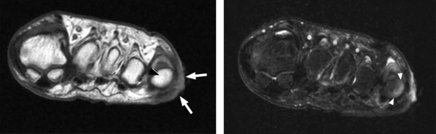
Short-axis T1 and fat-suppressed T2 MRI of the forefoot with arrows at a lateral ulcer and arrowheads at the fifth metatarsal head
Early osteomyelitis of the fifth metatarsal head beside a lateral ulcer (arrows), 54-year-old woman with diabetes. Short-axis T1 (left): marrow fat is still preserved (arrowhead). Fat-suppressed T2 (right): subcortical marrow edema (arrowheads). Next to an ulcer this pattern still means a high likelihood of early infection; confluent low T1 signal is the more specific sign. Image: Alaia EF, Chhabra A, Simpfendorfer CS, et al. MRI nomenclature for musculoskeletal infection. Skeletal Radiol 2021;50:2319-2347 (Fig. 16). CC BY 4.0. Changes: cropped; panel letters removed; arrows and arrowheads from the original figure.

Osteomyelitis or neuropathic (Charcot) joint?

Feature Osteomyelitis Neuropathic arthropathy
Location Under an ulcer: toes, metatarsal heads, calcaneus Midfoot (tarsometatarsal, intertarsal joints)
Ulcer and sinus tract Present Absent
Joints Usually one bone Several joints
Marrow Confluent low T1 under the ulcer Subchondral edema around joints, T1 often preserved
Cortex Disrupted Intact

Both show abnormal marrow signal. Osteomyelitis can arise in a Charcot foot when an ulcer forms over it.

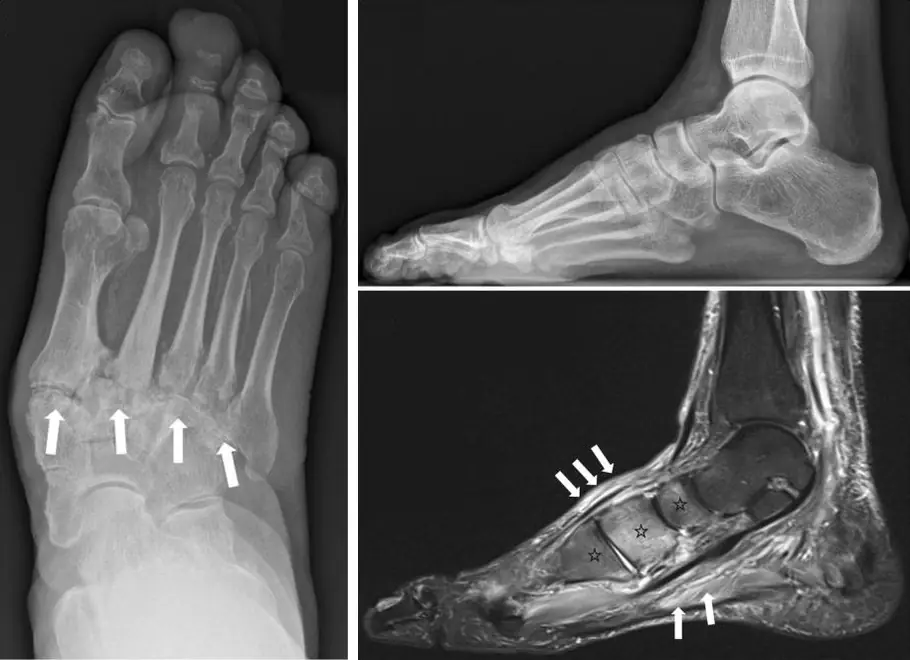
Dorsoplantar foot radiograph with arrows along the tarsometatarsal joints, a normal lateral radiograph and sagittal STIR MRI with midfoot marrow edema
Neuropathic (Charcot) midfoot, two patients. Left, dorsoplantar radiograph: the tarsometatarsal (Lisfranc) joints are involved, with lateral subluxation of the metatarsals (arrows). Right, early active Charcot foot: the lateral radiograph (top) is still normal, while sagittal STIR (bottom) shows marrow edema in several midfoot bones around their joints (asterisks) and soft-tissue edema (arrows), with no ulcer or sinus tract. Image: Rosskopf AB, Loupatatzis C, Pfirrmann CWA, Böni T, Berli MC. The Charcot foot: a pictorial review. Insights Imaging 2019;10:77 (Figs. 7a and 14). CC BY 4.0. Changes: cropped (two figures combined); panel letters and side marker removed; arrows and asterisks from the original figures.
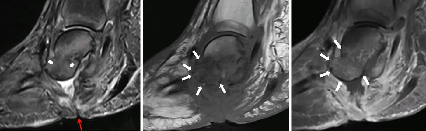
Three sagittal MRI images of a Charcot hindfoot with a red arrow at a skin ulcer and white arrows around the talus
Charcot foot complicated by osteomyelitis. Left, fat-suppressed fluid-sensitive image: a skin ulcer and sinus tract run from the sole up to the talus (red arrow). Middle, T1: diffuse marrow change in the talus, whose outline fades away (arrows). Right, post-contrast fat-suppressed T1: the outline reappears as it enhances (arrows), the 'ghost sign' described in the infected Charcot foot. Image: Rosskopf AB, Loupatatzis C, Pfirrmann CWA, Böni T, Berli MC. The Charcot foot: a pictorial review. Insights Imaging 2019;10:77 (Fig. 21). CC BY 4.0. Changes: cropped; panel letters removed; arrows from the original figure.

Further reading

  1. Ahmadi ME, Morrison WB, Carrino JA, et al. Neuropathic Arthropathy of the Foot with and without Superimposed Osteomyelitis: MR Imaging Characteristics. Radiology. 2006;238(2):622-631. doi:10.1148/radiol.2382041393
  2. Martín-Noguerol T, Luna Alcalá A, Beltrán LS, et al. Advanced MR Imaging Techniques for Differentiation of Neuropathic Arthropathy and Osteomyelitis in the Diabetic Foot. RadioGraphics. 2017;37(4):1161-1180. doi:10.1148/rg.2017160101

Practice this in RadQBoard. Chapter 7 has 30 board-style questions with full explanations. Free with an account.

Open RadQBoard Chapter 7 overview

Written in original words by the RadQBoard team. Images are reused under open licenses, with the source, license and any changes under each one.