Musculoskeletal · Chapter 5: Cartilage, fibrous, vascular and marrow tumors of bone · Page 1 of 12 · Free to read
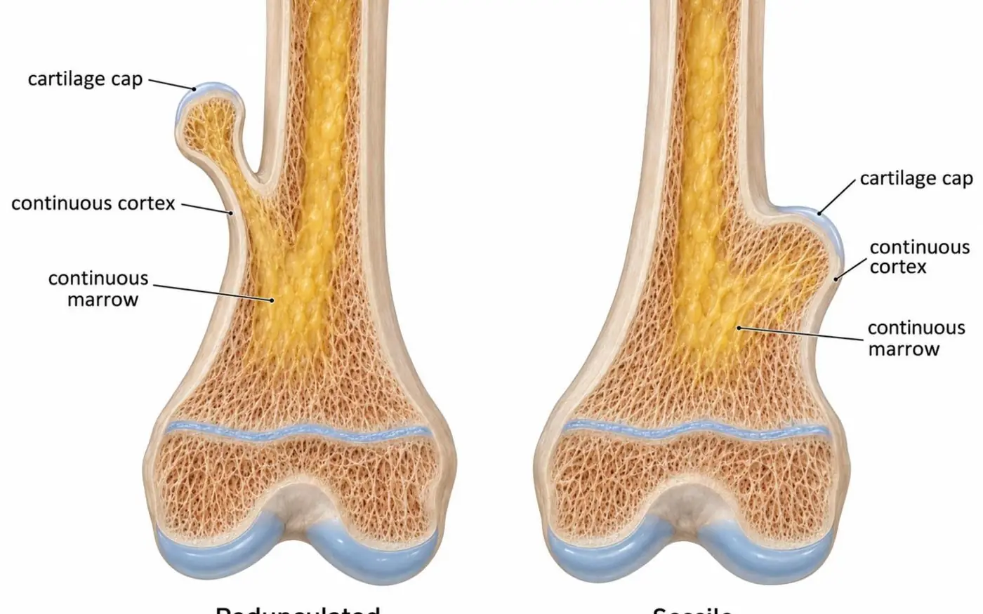
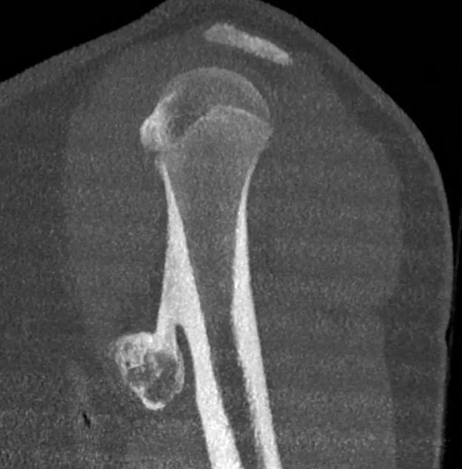
A bony outgrowth whose cortex and marrow run straight into the parent bone, pointing away from the joint.
What it is
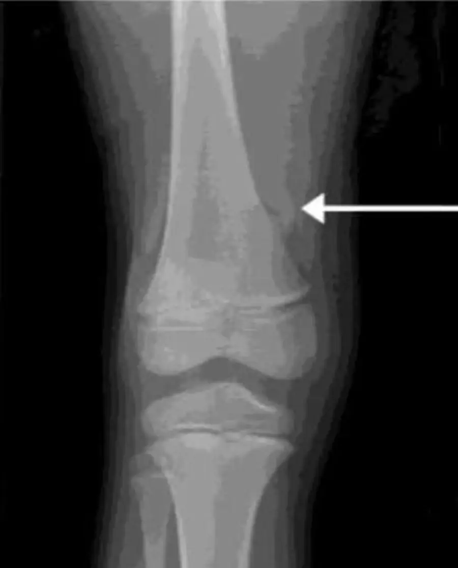
An osteochondroma is a cartilage-capped bony projection from the surface of a bone, either on a stalk (pedunculated) or broad-based (sessile). It is the most common benign bone lesion.
Children and adolescents; it often presents as a painless lump.
It grows with the skeleton and normally stops growing at skeletal maturity.
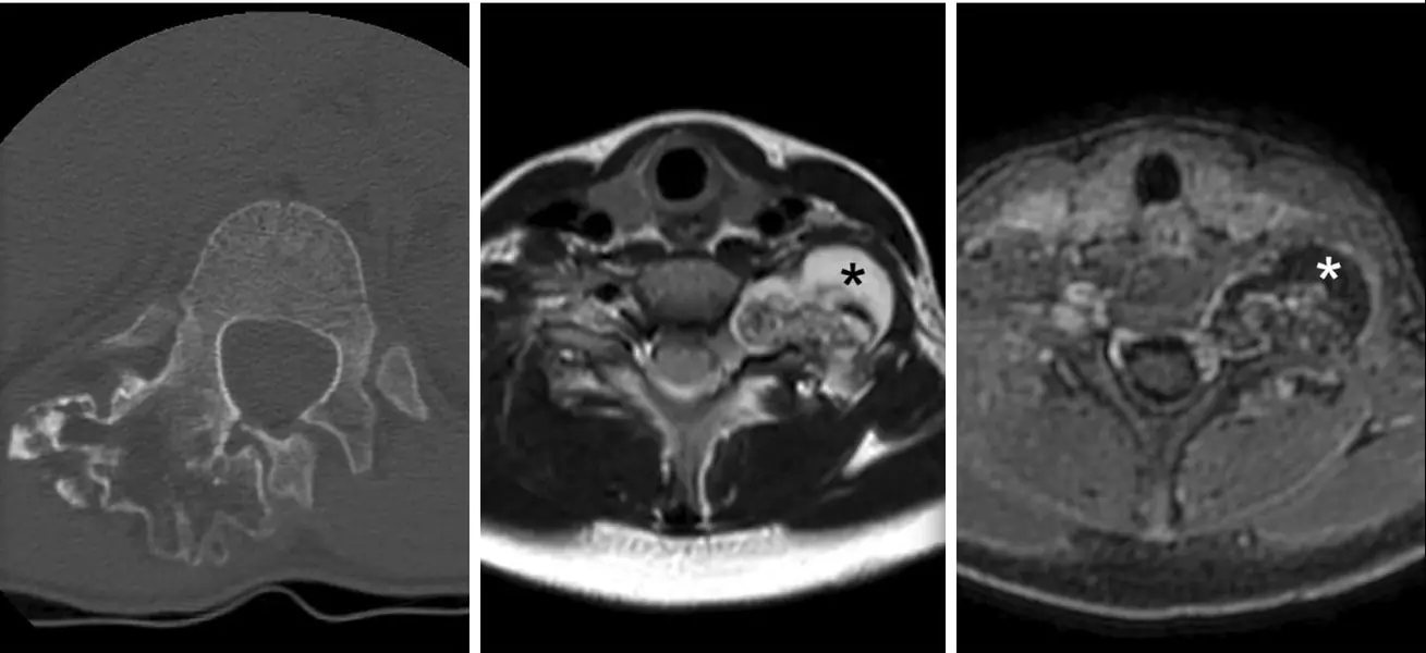
Measures the cartilage cap, bright on T2. A cap thicker than 2 cm on MRI suggests chondrosarcoma (many readers already worry above 1.5 cm in an adult).
Malignant transformation to chondrosarcoma (uncommon): pain without a fracture, growth after skeletal maturity, a thick cap or a soft-tissue mass.
Fracture of the stalk, an overlying bursa (bursitis), and compression of nerves or vessels, including pseudoaneurysm of the popliteal artery.
Multiple hereditary exostoses
An autosomal dominant condition (EXT1 or EXT2) with many osteochondromas, most often around the knees.
Limb deformities and short stature, such as a shortened ulna with bowing of the forearm.
A higher risk of chondrosarcoma than a solitary lesion.
Forearm lesions shorten the ulna and bow the radius (a Madelung-like deformity); the lifetime risk of malignant change is a few percent, higher than for a solitary lesion.
Murphey MD, Choi JJ, Kransdorf MJ, et al. Imaging of Osteochondroma: Variants and Complications with Radiologic-Pathologic Correlation. RadioGraphics. 2000;20(5):1407-1434. doi:10.1148/radiographics.20.5.g00se171407
Practice this in RadQBoard. Chapter 5 has 34 board-style questions with full explanations. Free with an account.