RadQBoard

RadQBoard › Musculoskeletal › Chapter 4 › Osteoid osteoma

Osteoid osteoma

Musculoskeletal · Chapter 4: Bone-forming and cartilage-forming tumors · Page 1 of 10 · Free to read

A tiny painful nidus wrapped in a lot of reactive bone: night pain that aspirin relieves, in a teenager.

What it is

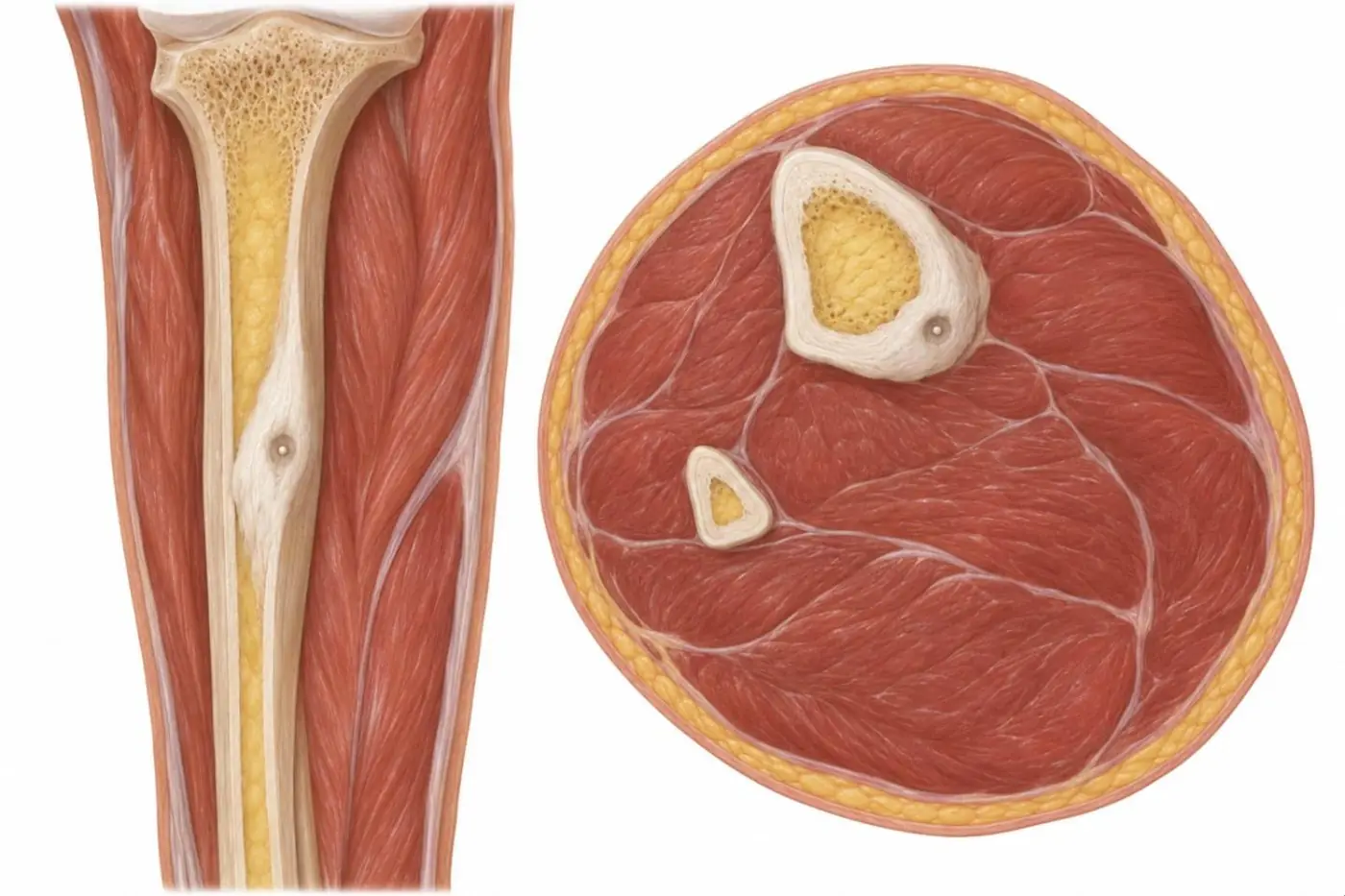
Osteoid osteoma is a small benign bone-forming tumor. Its core, the nidus, is a nest of osteoid and woven bone in a vascular stroma, usually smaller than 1.5–2 cm; around it the bone reacts with dense sclerosis.

Illustration of a tibial shaft with a spindle-shaped thick cortex holding a tiny round nidus, and a cross-section of the leg with the nidus in the thick tibial cortex
Illustration: osteoid osteoma of the tibial shaft. Left, longitudinal cut-away: a spindle-shaped thickening of the cortex from dense reactive bone, with a small round lucent nidus and a central dot of mineralization inside it. Right, cross-section of the leg at the same level: the nidus sits in the thickened posteromedial cortex of the tibia; the marrow cavity and the fibula are normal. Illustration adapted from CT in Diala PC, et al. Skeletal Radiol 2026 (Fig. 2) and a radiograph in Hanff DF, et al. J Belg Soc Radiol 2026 (Fig. 4) (CC BY 4.0). CC BY 4.0. Changes: simplified drawing of the pattern in the source figure; no tracing. Source articles: 1, 2.

Where it strikes

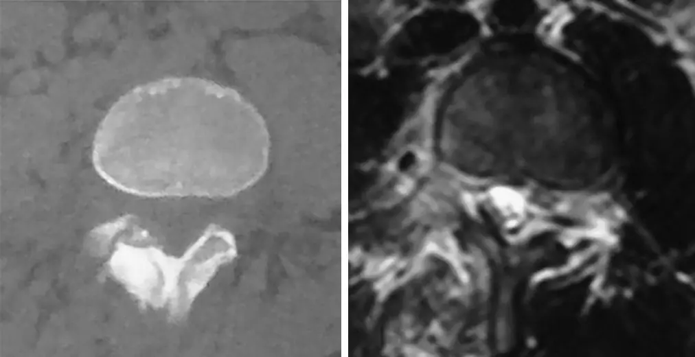
Axial CT and axial MRI of a vertebra with a small mineralized nidus in the lamina
Spinal osteoid osteoma in a 13-year-old boy with back pain and scoliosis. Left, axial CT: the nidus, with central mineralization, lies in the right lamina. Right, axial MRI: bright edema in the bone and soft tissue around the nidus. Treatment of the nidus allowed the scoliosis to correct. Image: Pan X, Qiao J, Liu Z, et al. Spontaneous correction of scoliosis after curettage of spinal osteoid osteoma: how and when? J Orthop Surg Res 2022;17:534 (Fig. 1c, d). CC BY 4.0. Changes: cropped (2 of 8 panels); scoliosis radiographs with handwritten measurements omitted; panel letters removed.
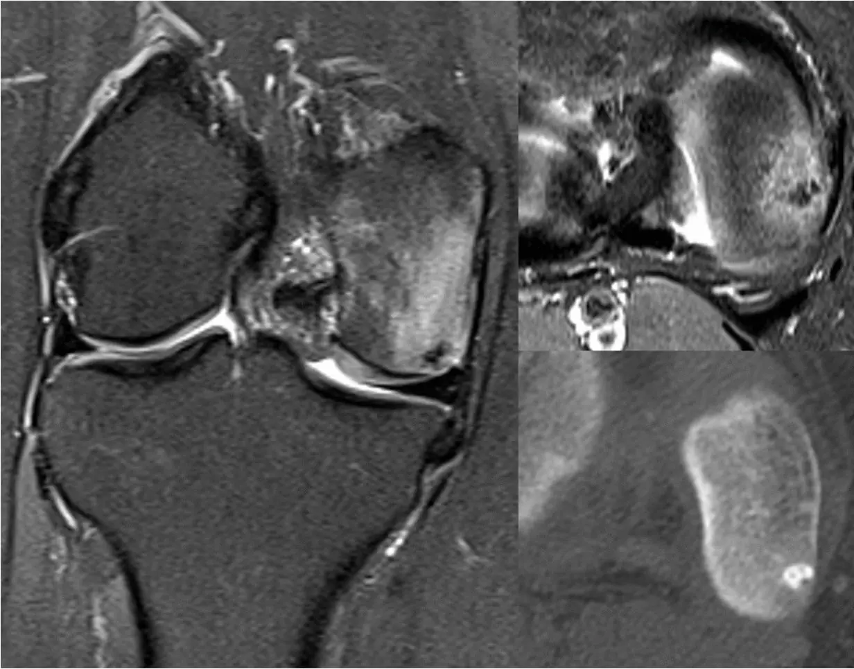
Coronal STIR MRI of the knee with edema in the medial femoral condyle, axial fat-suppressed MRI and axial CT with a small dense focus
Osteoid osteoma of the medial femoral condyle in a 24-year-old woman, near the joint. Left, coronal STIR: a small low-signal focus hidden in extensive marrow edema. Top right, axial proton-density fat-suppressed image: the same edema-like signal. Bottom right, axial CT: the subchondral nidus with its sclerotic core, shown far better than on MRI. Image: Wali L, Hegde G, Khoo M, et al. Intraosseous tumours of the knee. Skeletal Radiol 2026;55:1955-1982 (Fig. 6). CC BY 4.0. Changes: cropped; histology panel omitted.

Radiographs and CT

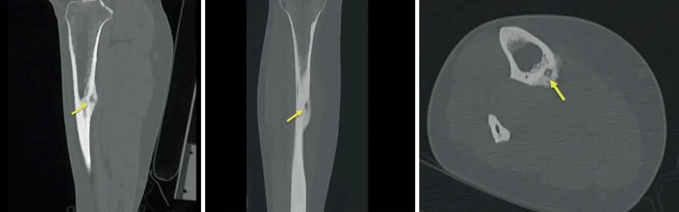
Sagittal, coronal and axial CT of the tibia with yellow arrows at a small lucent cortical lesion
Osteoid osteoma of the tibial shaft on CT: sagittal (left), coronal (middle) and axial (right). A subperiosteal nidus along the posteromedial mid-shaft (yellow arrows), with central calcification, cortical thickening and periosteal new bone. CT shows the nidus and its exact position for ablation. Image: Diala PC, Pluym I, Khan N, et al. CT-guided radiofrequency ablation of a tibial osteoid osteoma in pregnancy. Skeletal Radiol 2026;55:3001-3007 (Fig. 2). CC BY 4.0. Changes: cropped; panel letters cropped off; arrows from the original figure.

Bone scan

Intense uptake with the double density sign: a hot central focus (the nidus) inside a larger zone of milder uptake (the reactive sclerosis).

MRI

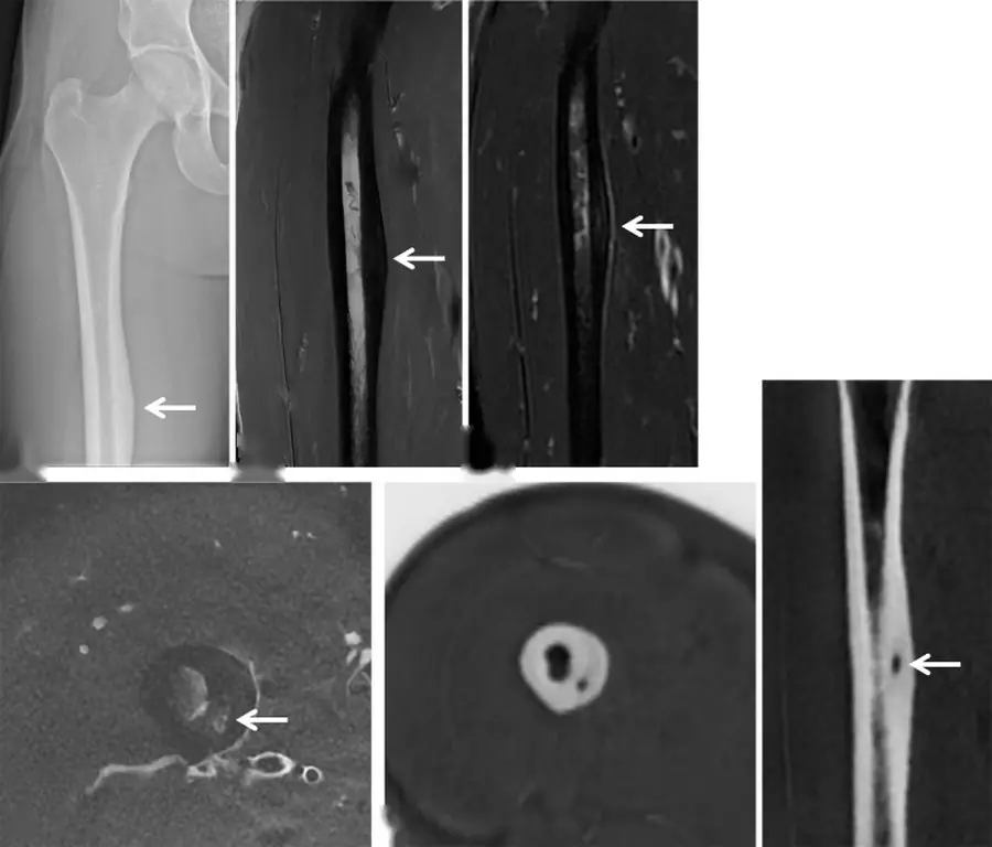
Radiograph of the femur, coronal and axial MRI and zero echo time MRI with arrows at a cortical nidus
Osteoid osteoma of the femoral shaft in a 23-year-old athlete. Top, from left: radiograph with focal thickening of the medial cortex (arrow); coronal T1-weighted and fat-suppressed T2-weighted MRI with cortical thickening and periosteal reaction (arrows). Bottom: on axial fat-suppressed T2 the nidus is only a subtle bright dot (arrow), while zero echo time (ZTE) MRI, a CT-like sequence, shows the small nidus inside the thick cortex clearly (axial and coronal, arrow). Image: Hanff DF, Vanhoenacker FM, Oei EHG. Zero echo time MRI for osseous assessment of sports-related pathology in athletes: a pictorial essay. J Belg Soc Radiol 2026;110:17 (Fig. 4). CC BY 4.0. Changes: cropped; panel letters removed; arrows from the original figure.

Differential diagnosis

Lesion Clue
Stress fracture A linear lucency running across the cortex, not a round nidus
Brodie abscess Larger lucency, often with a serpentine tract; no central nidus calcification
Osteoblastoma Larger than 2 cm, more expansile, less reactive sclerosis
Chronic osteomyelitis Sequestrum, sinus tract, clinical infection

Treatment

CT-guided radiofrequency ablation is the usual treatment; surgical curettage or resection are alternatives.

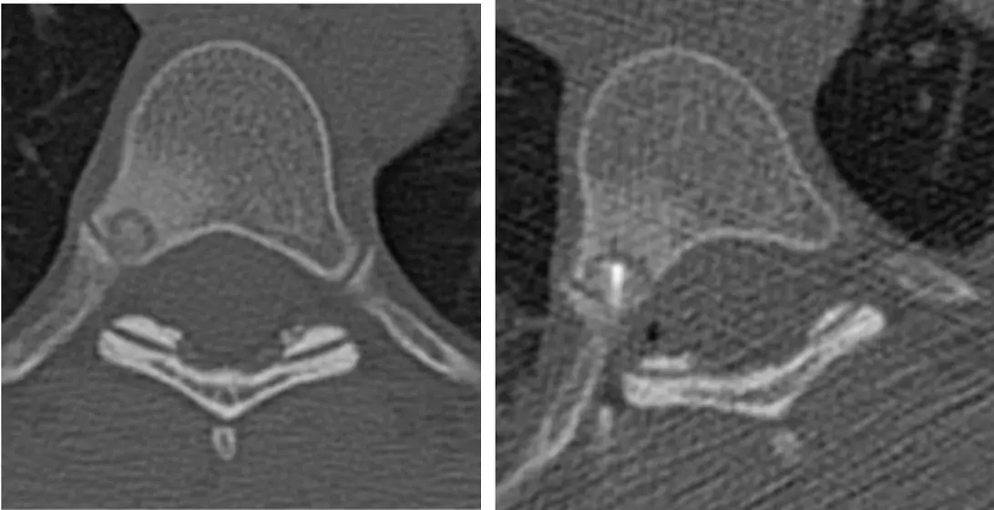
Two axial CT images of a thoracic vertebra, the second with an ablation probe in a small nidus
Osteoid osteoma of a thoracic vertebra treated with CT-guided radiofrequency ablation. Left, axial CT: a central sclerotic nidus with a lucent rim in the right posterior part of the vertebral body. Right: the ablation probe placed in the nidus. Image: Tregobov N, Krolikowski M, Dragoman R, et al. Unveiling primary bone tumors of the spine: a review of essential imaging clues. Diagnostics (Basel) 2025;15:2970 (Fig. 2b, c). CC BY 4.0. Changes: cropped (2 of 3 panels); panel letters cropped off.

Further reading

  1. Chai JW, Hong SH, Choi JY, et al. Radiologic Diagnosis of Osteoid Osteoma: From Simple to Challenging Findings. RadioGraphics. 2010;30(3):737-749. doi:10.1148/rg.303095120

Practice this in RadQBoard. Chapter 4 has 30 board-style questions with full explanations. Free with an account.
Open RadQBoard Chapter 4 overview

Written in original words by the RadQBoard team. Images are reused under open licenses, with the source, license and any changes under each one.