RadQBoard › Musculoskeletal › Chapter 3 › Periosteal reaction
Periosteal reaction
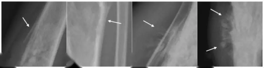
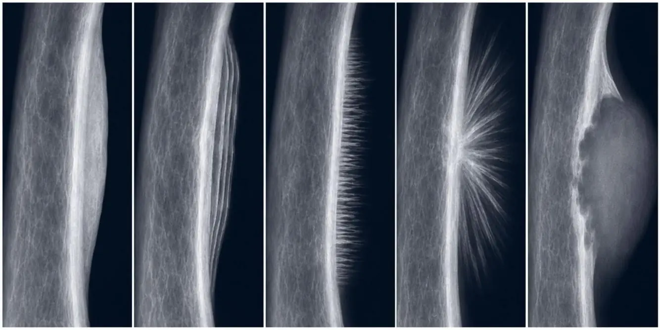
The faster a lesion grows, the less time the periosteum has to lay down bone, and the more broken the new bone looks.
What it is
The periosteum covers the outer surface of bone everywhere except the joint surfaces. When lifted by tumor, pus, blood or a healing process, its cells lay down new bone. The shape of that new bone tells how fast the process beneath it is growing.
Patterns, from slow to fast
| Pattern | Looks like | Growth | Examples |
|---|---|---|---|
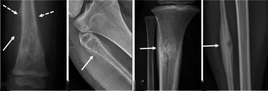
| Solid | Thick, smooth, continuous new bone | Slow | Osteoid osteoma, healing fracture, chronic osteomyelitis |
| Lamellated (onion skin) | Parallel layers | Stop-and-start | Ewing sarcoma, osteomyelitis, Langerhans cell histiocytosis |
| Spiculated: hair-on-end | Spicules perpendicular to the cortex | Fast | Ewing sarcoma |
| Spiculated: sunburst | Spicules radiating from one point | Very fast | Osteosarcoma |
| Codman triangle | New bone only at the edge where the periosteum is lifted | Fastest | Osteosarcoma, Ewing sarcoma; also abscess or hematoma |
Caveats
- Aggressive-looking periosteal reaction is not always tumor: infection, a healing stress fracture and Langerhans cell histiocytosis can do it.
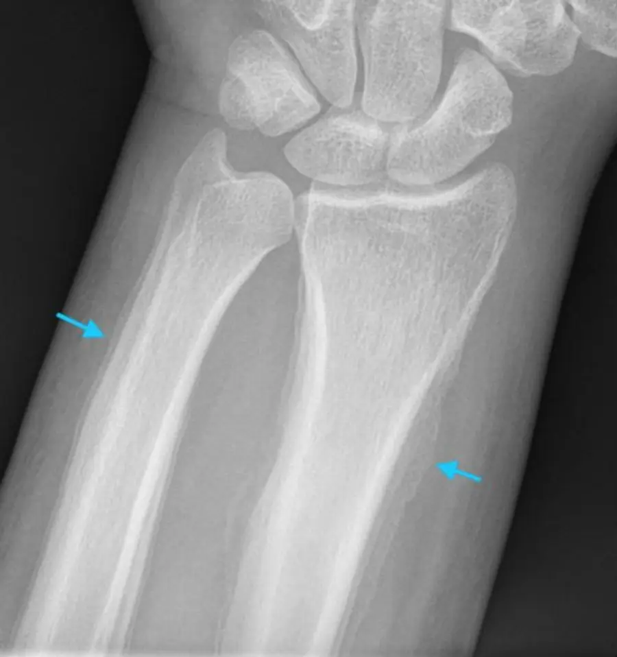
- Smooth, symmetric periosteal reaction of many long bones is usually systemic, not a tumor: hypertrophic osteoarthropathy (look for a lung tumor), venous stasis, thyroid acropachy, prostaglandin therapy and physiologic new bone in infants.
- Symmetric periosteal reaction of the long bones can be physiologic in the first months of life. In adults, think of hypertrophic osteoarthropathy, venous stasis and thyroid acropachy.
Further reading
- Rana RS, Wu JS, Eisenberg RL. Periosteal Reaction. American Journal of Roentgenology. 2009;193(4):W259-W272. doi:10.2214/ajr.09.3300
Practice this in RadQBoard. Chapter 3 has 32 board-style questions with full explanations. Free with an account.
Written in original words by the RadQBoard team. Images are reused under open licenses, with the source, license and any changes under each one.