RadQBoard › Musculoskeletal › Chapter 3
Metabolic arthropathies, neuropathic joints and the approach to bone tumors
The first half covers joint diseases driven by deposits, bleeding, growth or lost sensation: hemochromatosis, amyloid, ochronosis, sarcoidosis, hemophilia, juvenile idiopathic arthritis and the neuropathic joint. The second half sets out a working approach to any bone lesion, reading periosteal reaction, margins, matrix, age and location, and ends with quiet bone-forming lesions such as the bone island.
Free page: Periosteal reaction
Read all 10 pages and answer the chapter's 32 questions in the app. Free with an account.
What this chapter covers
Hemochromatosis and acromegaly
Two metabolic diseases that leave beak-shaped osteophytes on the metacarpal heads: iron narrows the joint, growth hormone first widens it.
Amyloid arthropathy and ochronosis
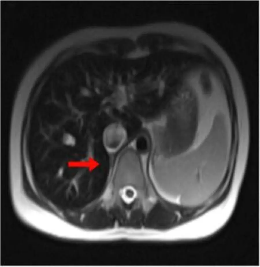
Two deposition diseases: amyloid lines the joints with lumpy, dark-on-T2 tissue; ochronosis calcifies every disc.
Sarcoidosis and multicentric reticulohistiocytosis
Lace in the phalanges means sarcoid; skin nodules with sharply punched-out DIP erosions mean multicentric reticulohistiocytosis.
Hemophilic arthropathy
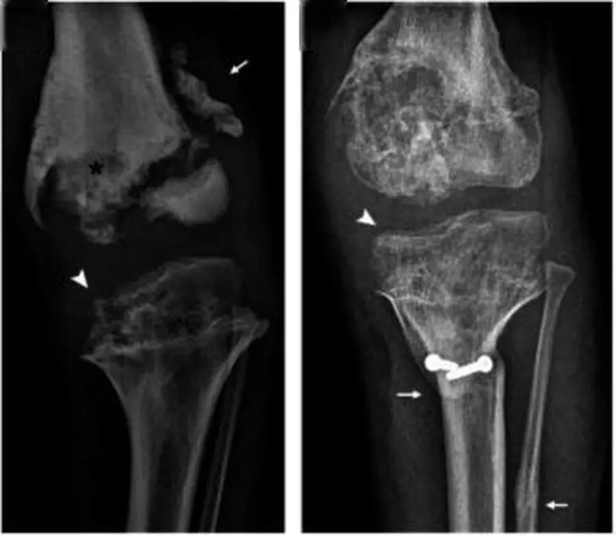
Repeated bleeding into a growing joint makes the bone ends big, the joint narrow and the synovium dense with iron.
Juvenile idiopathic arthritis
Inflammation in a growing skeleton changes the size and shape of the bones, and the joints can fuse.
Neuropathic (Charcot) joint
A joint without feeling keeps working until it falls apart: destruction, dislocation, debris and disorganization, with bone density preserved.
Bone tumors: the approach
First decide whether the lesion looks aggressive; then ask whether it has a feature that names it.
Periosteal reactionFree
The faster a lesion grows, the less time the periosteum has to lay down bone, and the more broken the new bone looks.
Margins, matrix, age and location
The edge of a lesion shows its speed, the matrix shows what it makes, and age and location narrow the list.
Bone-forming lesions: bone island, osteoma, melorheostosis
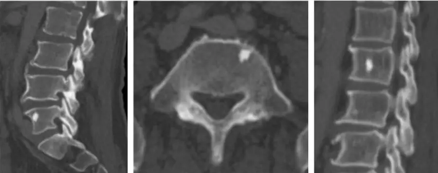
Dense bone that sits quietly: islands inside the marrow, osteomas on the cortex, and candle wax dripping along one limb.
Further reading
- Hirsch JH, Killien FC, Troupin RH. The Arthropathy of Hemochromatosis. Radiology. 1976;118(3):591-596. doi:10.1148/118.3.591
- Moore SL, Teirstein AE. Musculoskeletal Sarcoidosis: Spectrum of Appearances at MR Imaging. RadioGraphics. 2003;23(6):1389-1399. doi:10.1148/rg.236025172
- Sheybani EF, Khanna G, White AJ, et al. Imaging of Juvenile Idiopathic Arthritis: A Multimodality Approach. RadioGraphics. 2013;33(5):1253-1273. doi:10.1148/rg.335125178
- Jones EA, Manaster BJ, May DA, et al. Neuropathic Osteoarthropathy: Diagnostic Dilemmas and Differential Diagnosis. RadioGraphics. 2000;20(suppl_1):S279-S293. doi:10.1148/radiographics.20.suppl_1.g00oc22s279
- Miller TT. Bone Tumors and Tumorlike Conditions: Analysis with Conventional Radiography. Radiology. 2008;246(3):662-674. doi:10.1148/radiol.2463061038
- Rana RS, Wu JS, Eisenberg RL. Periosteal Reaction. American Journal of Roentgenology. 2009;193(4):W259-W272. doi:10.2214/ajr.09.3300
- Ihde LL, Forrester DM, Gottsegen CJ, et al. Sclerosing Bone Dysplasias: Review and Differentiation from Other Causes of Osteosclerosis. RadioGraphics. 2011;31(7):1865-1882. doi:10.1148/rg.317115093
Read all 10 pages and answer the chapter's 32 questions in the app. Free with an account.