RadQBoard › Musculoskeletal › Chapter 4
Bone-forming and cartilage-forming tumors
Bone-forming tumors run from the small painful nidus of osteoid osteoma to osteosarcoma in its conventional, telangiectatic and surface forms, including how it spreads. Cartilage-forming lesions follow: synovial chondromatosis, enchondroma, the features that should raise concern for chondrosarcoma, and the syndromes with many enchondromas.
Read all 10 pages and answer the chapter's 30 questions in the app. Free with an account.
What this chapter covers
Osteoid osteomaFree
A tiny painful nidus wrapped in a lot of reactive bone: night pain that aspirin relieves, in a teenager.
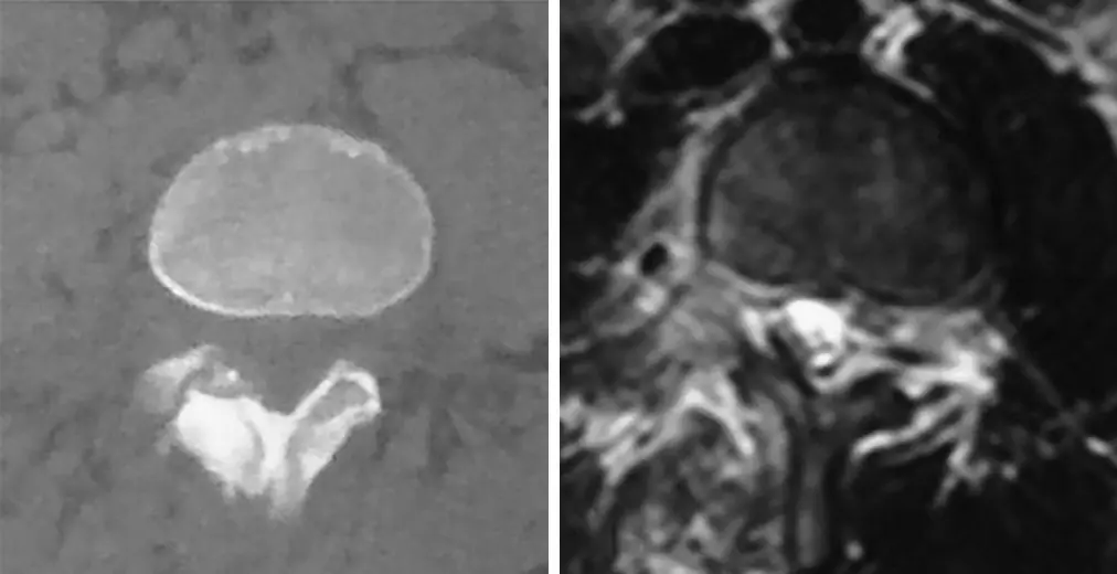
Osteoblastoma
The big brother of osteoid osteoma: same tissue, larger than 2 cm, and at home in the posterior elements of the spine.
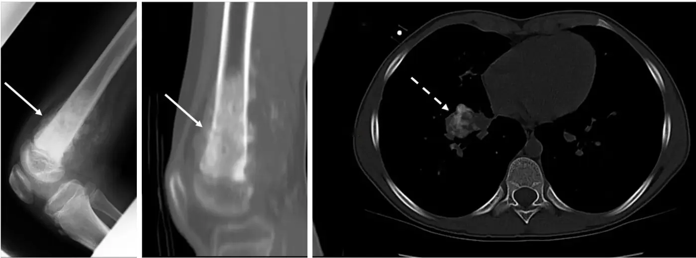
Osteosarcoma: the disease and the conventional type
Bone destruction, cloud-like new bone made by the tumor, a sunburst of periosteal reaction and a soft-tissue mass, around the knee of a teenager.
Telangiectatic osteosarcoma
A malignant tumor that looks like a blood-filled cyst: fluid-fluid levels, but with thick solid nodules that an aneurysmal bone cyst does not have.
Surface osteosarcomas: parosteal and periosteal
Parosteal is the outer layer: an older patient, a dense cauliflower behind the femur. Periosteal is the inner layer: a teenager, a shaft lesion with spiculated reaction.
Osteosarcoma spread and secondary osteosarcoma
Whatever the subtype, osteosarcoma goes to the lungs, and its metastases can make bone there too.
Synovial chondromatosis
Many small bodies of the same size inside one joint: the synovium is making cartilage.
Enchondroma
Rings and arcs in a long bone; a lucent, expanding lesion in a finger.
Enchondroma or chondrosarcoma?
Most enchondromas never change. Worry about new pain without a fracture, deepening scalloping and disappearing calcification.
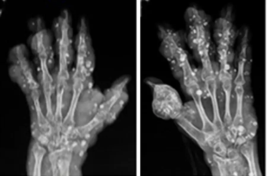
Ollier disease and Maffucci syndrome
Many enchondromas: Ollier alone; with venous malformations and phleboliths, Maffucci.
Further reading
- Chai JW, Hong SH, Choi JY, et al. Radiologic Diagnosis of Osteoid Osteoma: From Simple to Challenging Findings. RadioGraphics. 2010;30(3):737-749. doi:10.1148/rg.303095120
- Murphey MD, Robbin MR, McRae GA, et al. The many faces of osteosarcoma. RadioGraphics. 1997;17(5):1205-1231. doi:10.1148/radiographics.17.5.9308111
- Murphey MD, wan Jaovisidha S, Temple HT, et al. Telangiectatic Osteosarcoma: Radiologic-Pathologic Comparison. Radiology. 2003;229(2):545-553. doi:10.1148/radiol.2292021130
- Murphey MD, Jelinek JS, Temple HT, et al. Imaging of Periosteal Osteosarcoma: Radiologic-Pathologic Comparison. Radiology. 2004;233(1):129-138. doi:10.1148/radiol.2331030326
- Jelinek JS, Murphey MD, Kransdorf MJ, et al. Parosteal osteosarcoma: value of MR imaging and CT in the prediction of histologic grade. Radiology. 1996;201(3):837-842. doi:10.1148/radiology.201.3.8939240
- Murphey MD, Vidal JA, Fanburg-Smith JC, et al. Imaging of Synovial Chondromatosis with Radiologic-Pathologic Correlation. RadioGraphics. 2007;27(5):1465-1488. doi:10.1148/rg.275075116
- Murphey MD, Flemming DJ, Boyea SR, et al. Enchondroma versus chondrosarcoma in the appendicular skeleton: differentiating features. RadioGraphics. 1998;18(5):1213-1237. doi:10.1148/radiographics.18.5.9747616
Read all 10 pages and answer the chapter's 30 questions in the app. Free with an account.