RadQBoard › Musculoskeletal › Chapter 5
Cartilage, fibrous, vascular and marrow tumors of bone
This chapter finishes the bone tumors: cartilage lesions from osteochondroma to chondrosarcoma, fibrous lesions such as nonossifying fibroma and fibrous dysplasia, vertebral hemangioma and giant cell tumor. It closes with Langerhans cell histiocytosis and the round cell and marrow tumors, Ewing sarcoma, myeloma and lymphoma, plus intraosseous lipoma and chordoma.
Free page: Osteochondroma and hereditary exostoses
Read all 12 pages and answer the chapter's 34 questions in the app. Free with an account.
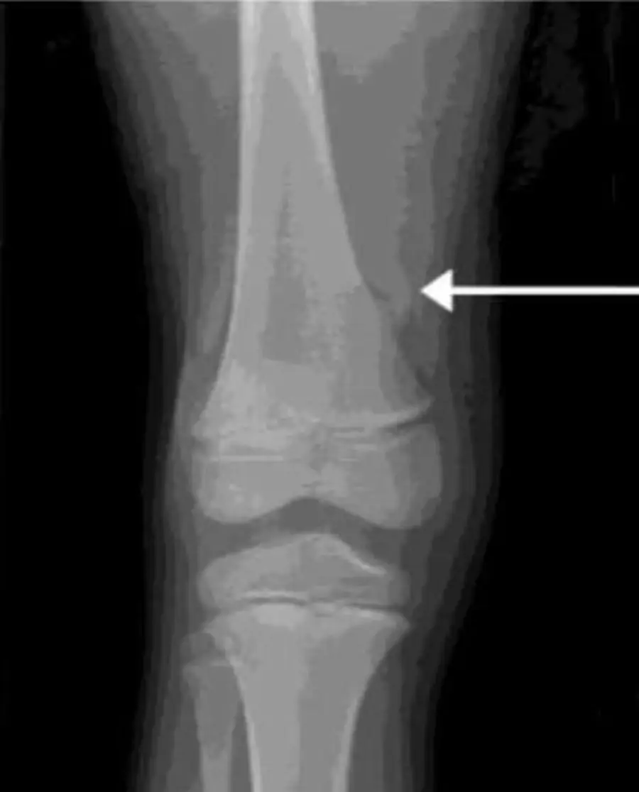
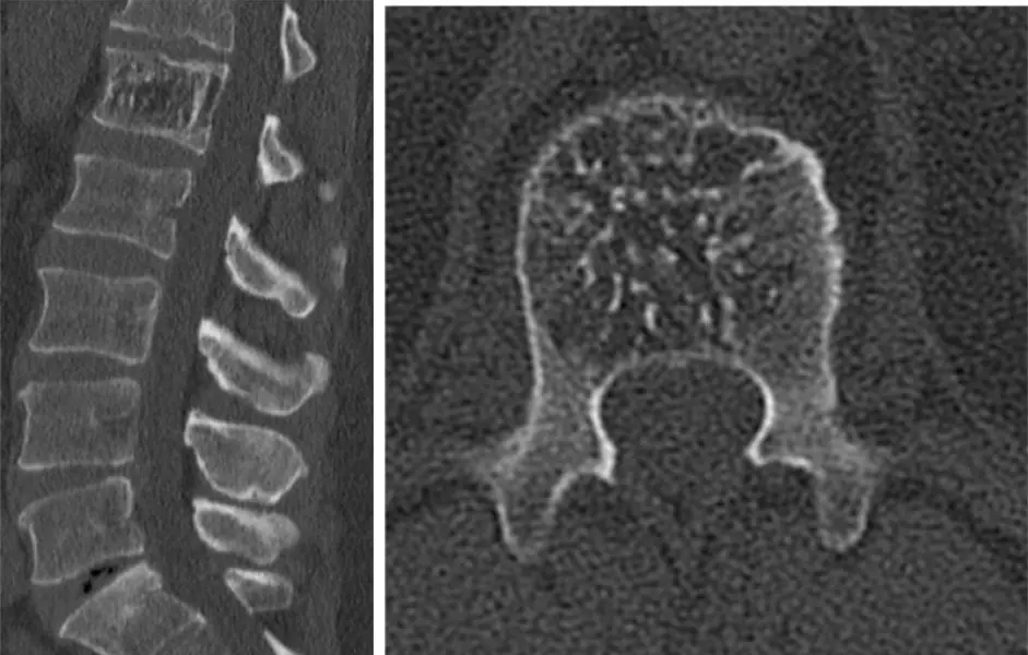
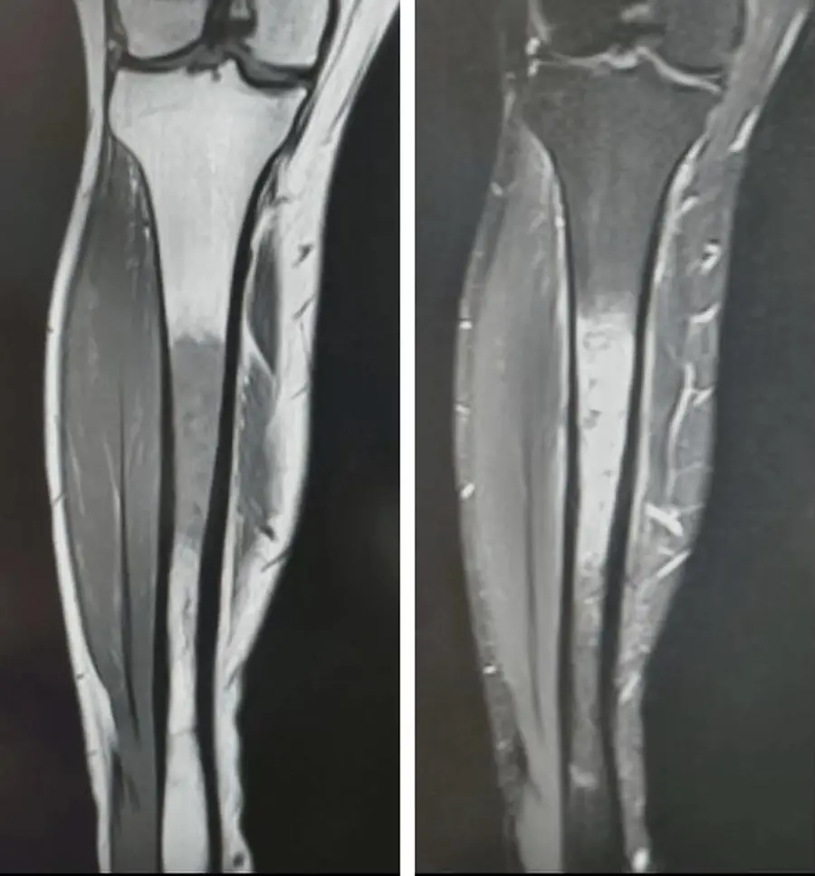
What this chapter covers
Osteochondroma and hereditary exostosesFree
A bony outgrowth whose cortex and marrow run straight into the parent bone, pointing away from the joint.
Chondroblastoma and chondromyxoid fibroma
A lucent lesion in the epiphysis of a child is chondroblastoma until proven otherwise, and it is the cartilage lesion that is dark on T2.
Chondrosarcoma
An adult with a painful cartilage lesion in the pelvis or proximal femur: rings and arcs, deep endosteal scalloping and a soft-tissue mass.
Nonossifying fibroma and fibrous cortical defect
A bubbly lucent lesion in the cortex of a child's knee, with a sclerotic scalloped rim: leave it alone.
Fibrous dysplasia in the long bones and pelvis
Ground-glass bone in a young patient, sometimes bending the femur into a shepherd's crook.
Fibrous dysplasia of the skull and its syndromes
An expanded skull base with ground-glass bone: fibrous dysplasia in the young, Paget disease in the old.
Vertebral hemangioma
Coarse vertical stripes in a vertebral body, polka dots on the axial image, and bright on T1 and T2 from fat.
Giant cell tumor of bone
An adult with closed growth plates and an eccentric lucent lesion that runs up to the articular surface, with no sclerotic rim.
Langerhans cell histiocytosis of bone
The great mimicker of childhood: a beveled hole in the skull, a flattened vertebra, or an aggressive lesion in a long bone.
Ewing sarcoma
A child with pain and fever, a permeative lesion in the shaft and a big soft-tissue mass: it looks like infection, and it is a sarcoma.
Multiple myeloma and plasmacytoma
Many punched-out holes in an adult over 40, often cold on bone scan.
Lymphoma, intraosseous lipoma and chordoma
Three last tumors with clues of their own: a soft-tissue mass with little bone destruction, fat with a central calcification, and a midline lobulated T2-bright mass.
Further reading
- Murphey MD, Choi JJ, Kransdorf MJ, et al. Imaging of Osteochondroma: Variants and Complications with Radiologic-Pathologic Correlation. RadioGraphics. 2000;20(5):1407-1434. doi:10.1148/radiographics.20.5.g00se171407
- Murphey MD, Walker EA, Wilson AJ, et al. From the Archives of the AFIP: Imaging of Primary Chondrosarcoma: Radiologic-Pathologic Correlation. RadioGraphics. 2003;23(5):1245-1278. doi:10.1148/rg.235035134
- Kushchayeva YS, Kushchayev SV, Glushko TY, et al. Fibrous dysplasia for radiologists: beyond ground glass bone matrix. Insights into Imaging. 2018;9(6):1035-1056. doi:10.1007/s13244-018-0666-6 · Free full text
- Murphey MD, Nomikos GC, Flemming DJ, et al. Imaging of Giant Cell Tumor and Giant Cell Reparative Granuloma of Bone: Radiologic-Pathologic Correlation. RadioGraphics. 2001;21(5):1283-1309. doi:10.1148/radiographics.21.5.g01se251283
- Stull MA, Kransdorf MJ, Devaney KO. Langerhans cell histiocytosis of bone. RadioGraphics. 1992;12(4):801-823. doi:10.1148/radiographics.12.4.1636041
- Murphey MD, Senchak LT, Mambalam PK, et al. From the Radiologic Pathology Archives: Ewing Sarcoma Family of Tumors: Radiologic-Pathologic Correlation. RadioGraphics. 2013;33(3):803-831. doi:10.1148/rg.333135005
- Ormond Filho AG, Carneiro BC, Pastore D, et al. Whole-Body Imaging of Multiple Myeloma: Diagnostic Criteria. RadioGraphics. 2019;39(4):1077-1097. doi:10.1148/rg.2019180096
Read all 12 pages and answer the chapter's 34 questions in the app. Free with an account.