RadQBoard › Musculoskeletal › Chapter 6
Bone cysts, metastases, tumor mimics and soft-tissue tumors
Cystic bone lesions and metastases open the chapter, followed by lesions that imitate tumors: myositis ossificans, brown tumor and cortical desmoid. The rest turns to soft tissue: fatty tumors from lipoma to liposarcoma, fibrolipomatous hamartoma of nerve, high-grade sarcoma, and fibrous lesions such as fibromatosis, elastofibroma, desmoid and DFSP.
Read all 11 pages and answer the chapter's 30 questions in the app. Free with an account.
What this chapter covers
Simple bone cyst
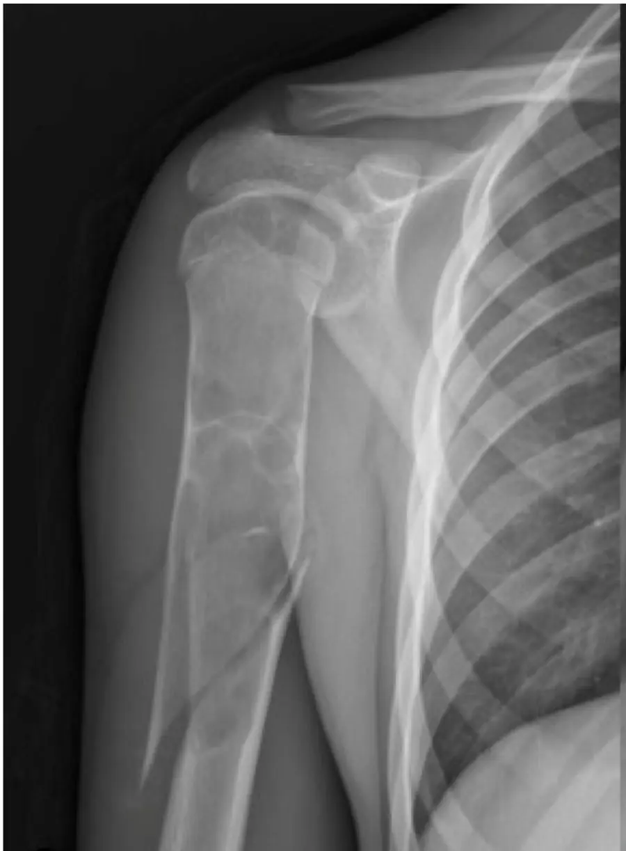
A central, fluid-filled lucency in the proximal humerus of a child, often found after it breaks, with a bone fragment lying in the bottom of the cyst.
Aneurysmal bone cyst and adamantinoma
A blown-out, multicystic lesion full of fluid-fluid levels in a teenager; if the biopsy says ABC, make sure nothing worse was hiding inside.
Bone metastases
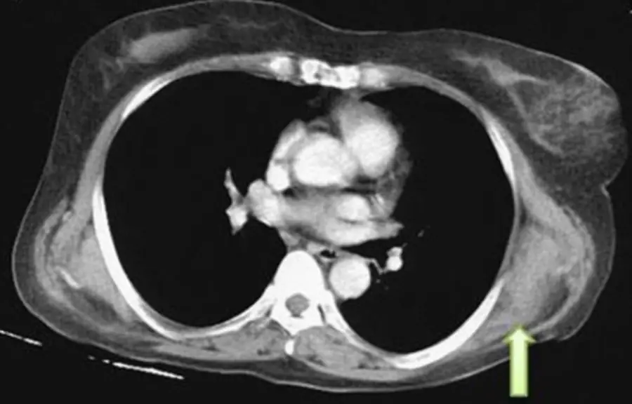
Ten times more common than primary bone tumors: an older patient with a lytic or sclerotic lesion has a metastasis until proven otherwise.
Myositis ossificans
Heterotopic bone in muscle after (sometimes forgotten) trauma: it matures from the outside in, and it must not be biopsied.
Brown tumor and cortical desmoid
Two benign mimics: a lytic lesion that only makes sense with the rest of hyperparathyroidism, and a ragged cortex behind a teenager's knee that should be left alone.
Lipoma and atypical lipomatous tumor
Fat that behaves like fat on every sequence is a lipoma; thick septa, nodules or enhancement mean atypical lipomatous tumor.
High-grade and myxoid liposarcoma
High-grade liposarcomas may show little or no fat; myxoid liposarcoma is bright on T2 like a cyst but enhances.
Fibrolipomatous hamartoma of nerve
A median nerve swollen with fat between its fascicles: a coaxial cable on axial images, spaghetti on long-axis images.
High-grade soft-tissue sarcoma
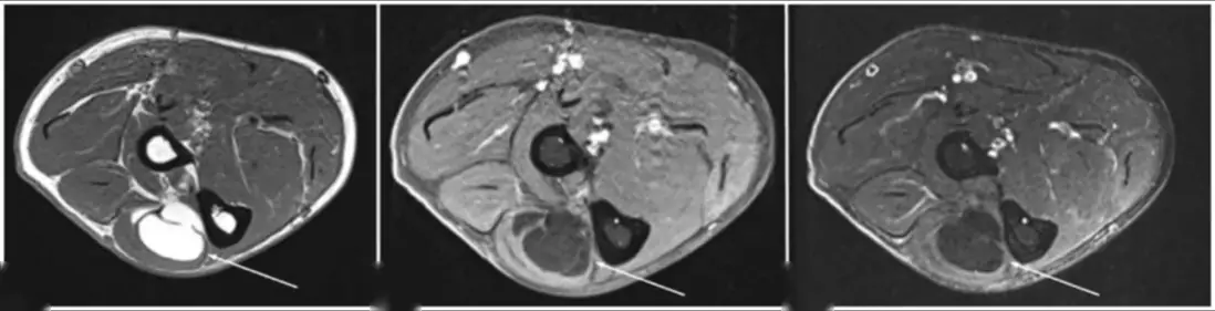
A large, deep, heterogeneous mass with necrosis in an adult's thigh: imaging says high-grade sarcoma; only pathology says which one.
Superficial fibromatosis and nodular fasciitis
Nodules along the palmar or plantar aponeurosis are fibromatosis; a fast-growing, enhancing mass on the fascia of a young adult may be nodular fasciitis.
Elastofibroma, desmoid and DFSP
A striped crescent under the scapula, a dark infiltrating band in the abdominal wall, and an enhancing skin nodule: three fibrous masses with three different futures.
Further reading
- Restrepo R, Zahrah D, Pelaez L, et al. Update on aneurysmal bone cyst: pathophysiology, histology, imaging and treatment. Pediatric Radiology. 2022;52(9):1601-1614. doi:10.1007/s00247-022-05396-6 · Free full text
- Kransdorf MJ, Sweet DE. Aneurysmal bone cyst: concept, controversy, clinical presentation, and imaging. American Journal of Roentgenology. 1995;164(3):573-580. doi:10.2214/ajr.164.3.7863874
- Lecouvet FE. Whole-Body MR Imaging: Musculoskeletal Applications. Radiology. 2016;279(2):345-365. doi:10.1148/radiol.2016142084
- Kransdorf MJ, Meis JM, Jelinek JS. Myositis ossificans: MR appearance with radiologic-pathologic correlation. American Journal of Roentgenology. 1991;157(6):1243-1248. doi:10.2214/ajr.157.6.1950874
- Murphey MD, Carroll JF, Flemming DJ, et al. From the Archives of the AFIP: Benign Musculoskeletal Lipomatous Lesions. RadioGraphics. 2004;24(5):1433-1466. doi:10.1148/rg.245045120
- Murphey MD, Arcara LK, Fanburg-Smith J. Imaging of Musculoskeletal Liposarcoma with Radiologic-Pathologic Correlation. RadioGraphics. 2005;25(5):1371-1395. doi:10.1148/rg.255055106
- Marom EM, Helms CA. Fibrolipomatous hamartoma: pathognomonic on MR imaging. Skeletal Radiology. 1999;28(5):260-264. doi:10.1007/s002560050512
- Kransdorf MJ, Murphey MD. Imaging of Soft-Tissue Musculoskeletal Masses: Fundamental Concepts. RadioGraphics. 2016;36(6):1931-1948. doi:10.1148/rg.2016160084
- Murphey MD, Ruble CM, Tyszko SM, et al. Musculoskeletal Fibromatoses: Radiologic-Pathologic Correlation. RadioGraphics. 2009;29(7):2143-2183. doi:10.1148/rg.297095138
Read all 11 pages and answer the chapter's 30 questions in the app. Free with an account.