RadQBoard › Musculoskeletal › Chapter 7
Nerve and other soft-tissue tumors, joint and bone infection
Nerve sheath tumors lead the chapter, with the signs that separate benign from malignant, followed by myxoma, synovial sarcoma, Morton neuroma and a practical soft-tissue differential. The second half covers infection: septic arthritis, the vocabulary and routes of osteomyelitis, hematogenous infection in children, the diabetic foot and chronic infection.
Free page: Contiguous osteomyelitis and the diabetic foot
Read all 10 pages and answer the chapter's 30 questions in the app. Free with an account.
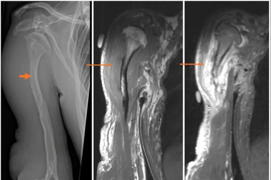
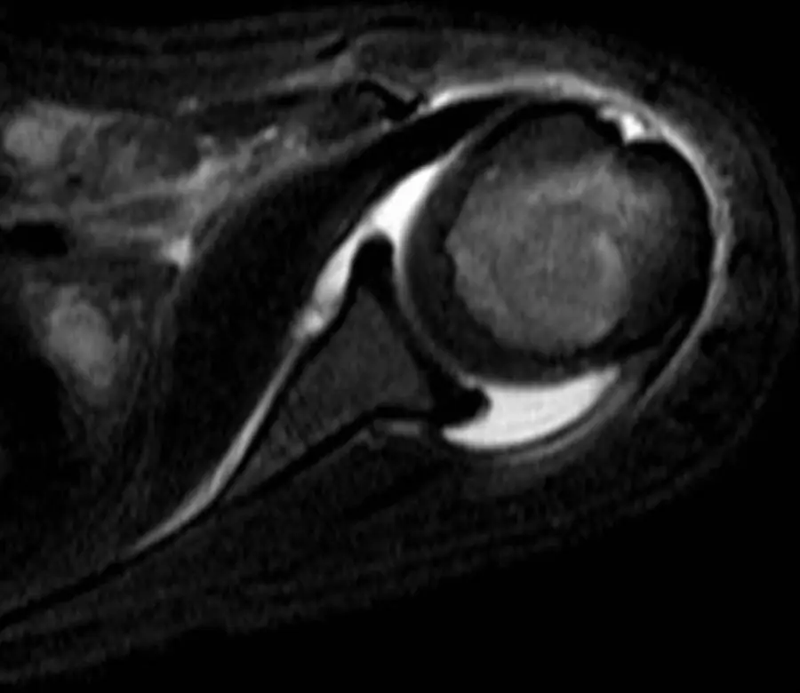
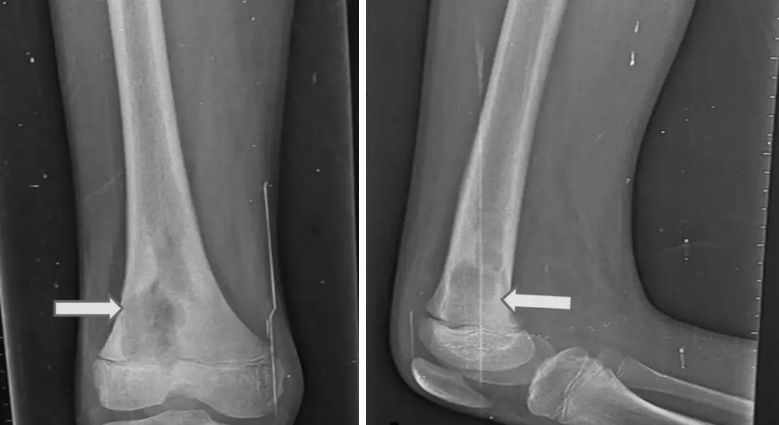
What this chapter covers
Schwannoma and neurofibroma
A fusiform, T2-bright, enhancing mass on the course of a nerve, with a cap of fat at each end and a target in the middle.
Malignant peripheral nerve sheath tumor
In a patient with neurofibromatosis type 1, a nerve tumor that grows, hurts or loses its target is malignant until proven otherwise.
Myxoma and synovial sarcoma
Two masses that look almost like fluid on T2: an intramuscular myxoma that barely enhances, and a synovial sarcoma near a joint in a young adult that does.
Morton neuroma
A dumbbell-shaped, dark-on-T2 mass between the metatarsal heads, below the ligament that joins them.
Soft-tissue tumor differential diagnosis
Most soft-tissue masses are indeterminate; a few announce themselves. Know the few, and send the rest for biopsy.
Septic arthritis
An emergency: one hot joint is infected until the aspirate says otherwise, and the radiograph is often normal.
Osteomyelitis: routes and vocabulary
Know the words (sequestrum, involucrum, cloaca, sinus tract, Brodie abscess) and how the infection got in.
Hematogenous osteomyelitis
A child's metaphysis, where slow blood lets bacteria settle; the infection starts inside and lifts the loose periosteum.
Contiguous osteomyelitis and the diabetic footFree
An ulcer over a bone with a track down to it is osteomyelitis; a swollen midfoot without an ulcer is usually Charcot joint.
Brodie abscess, chronic osteomyelitis and special cases
Subacute infection walls itself off; chronic infection leaves dead bone; some organisms and sites have their own stories.
Further reading
- Murphey MD, Smith WS, Smith SE, et al. From the Archives of the AFIP: Imaging of Musculoskeletal Neurogenic Tumors: Radiologic-Pathologic Correlation. RadioGraphics. 1999;19(5):1253-1280. doi:10.1148/radiographics.19.5.g99se101253
- Murphey MD, Gibson MS, Jennings BT, et al. Imaging of Synovial Sarcoma with Radiologic-Pathologic Correlation. RadioGraphics. 2006;26(5):1543-1565. doi:10.1148/rg.265065084
- Wu JS, Hochman MG. Soft-Tissue Tumors and Tumorlike Lesions: A Systematic Imaging Approach. Radiology. 2009;253(2):297-316. doi:10.1148/radiol.2532081199
- Chan BY, Crawford AM, Kobes PH, et al. Septic Arthritis: An Evidence-Based Review of Diagnosis and Image-Guided Aspiration. American Journal of Roentgenology. 2020;215(3):568-581. doi:10.2214/ajr.20.22773
- Jaramillo D, Dormans JP, Delgado J, et al. Hematogenous Osteomyelitis in Infants and Children: Imaging of a Changing Disease. Radiology. 2017;283(3):629-643. doi:10.1148/radiol.2017151929
- Ahmadi ME, Morrison WB, Carrino JA, et al. Neuropathic Arthropathy of the Foot with and without Superimposed Osteomyelitis: MR Imaging Characteristics. Radiology. 2006;238(2):622-631. doi:10.1148/radiol.2382041393
- Martín-Noguerol T, Luna Alcalá A, Beltrán LS, et al. Advanced MR Imaging Techniques for Differentiation of Neuropathic Arthropathy and Osteomyelitis in the Diabetic Foot. RadioGraphics. 2017;37(4):1161-1180. doi:10.1148/rg.2017160101
- Debs P, Boutin RD, Smith SE, et al. Chronic Nonspinal Osteomyelitis in Adults: Consensus Recommendations on Percutaneous Bone Biopsies from the Society of Academic Bone Radiologists. Radiology. 2024;311(1):e231348. doi:10.1148/radiol.231348
Read all 10 pages and answer the chapter's 30 questions in the app. Free with an account.