RadQBoard › Musculoskeletal › Chapter 8
Imaging infection and metabolic bone disease
The chapter compares radiographs, nuclear medicine and MRI for osteomyelitis and covers soft-tissue infections, including gas-forming infection. It then works through metabolic bone disease: osteoporosis and DEXA, scurvy and osteomalacia, endocrine bone disease, hyperparathyroidism, renal osteodystrophy, Paget disease and the rare sclerosing diseases.
Read all 10 pages and answer the chapter's 30 questions in the app. Free with an account.
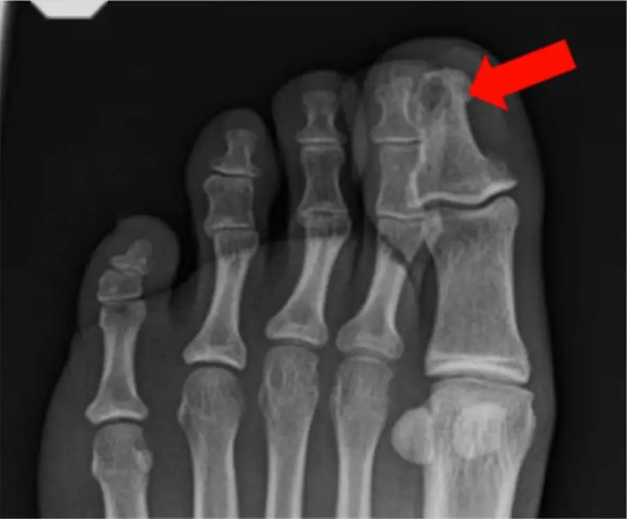
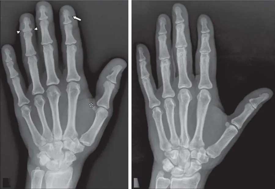
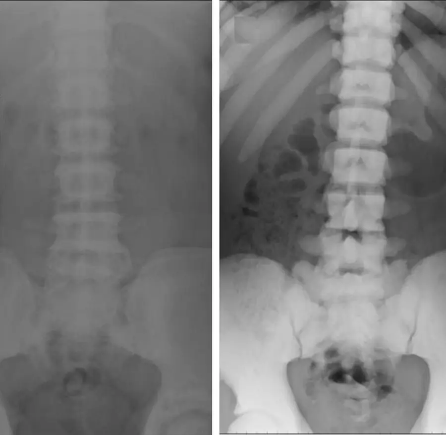
What this chapter covers
Imaging osteomyelitis: radiographs and nuclear medicine
Radiographs come first but lag by two weeks; bone scan is positive in a day or two but is not specific; adding white-cell imaging adds specificity.
MRI of osteomyelitis
Read the T1 images: confluent dark marrow in the medullary canal is osteomyelitis; hazy or thin subcortical change is not.
Soft-tissue infections
Gas in the soft tissues of a sick patient means necrotizing fasciitis until proven otherwise; an intramuscular abscess in a diabetic is pyomyositis.
Osteoporosis and DEXA
The most common metabolic bone disease; DEXA measures it, and the T-score defines it after menopause and over 50.
Scurvy and osteomalacia
Scurvy bleeds under the periosteum and rings the epiphysis; osteomalacia leaves bands of unmineralized bone across the cortex (Looser zones).
Acromegaly and thyroid bone disease
Too much growth hormone widens the hands, feet and face; the thyroid changes bone age, and treated Graves disease can make the fingers grow periosteal bone.
Hyperparathyroidism
Bone resorbed under the periosteum, the ligaments and the cartilage: look at the radial side of the middle phalanges, the distal clavicles and a salt-and-pepper skull.
Renal osteodystrophy and hypoparathyroidism
Kidney failure gives bones a rugger jersey; too little parathyroid hormone calcifies the basal ganglia; a short fourth metacarpal points to pseudohypoparathyroidism.
Paget diseaseFree
A caricature of normal bone in an older adult: bigger, thicker cortex, coarse trabeculae, and a lytic front shaped like a blade of grass.
Juvenile Paget disease, osteopetrosis and Gaucher disease
Three rare diseases with signature looks: a whole child's skeleton like Paget, a skeleton of chalk that breaks, and an Erlenmeyer flask femur with a big spleen.
Further reading
- Erdman WA, Tamburro F, Jayson HT, et al. Osteomyelitis: characteristics and pitfalls of diagnosis with MR imaging. Radiology. 1991;180(2):533-539. doi:10.1148/radiology.180.2.2068324
- Hayeri MR, Ziai P, Shehata ML, et al. Soft-Tissue Infections and Their Imaging Mimics: From Cellulitis to Necrotizing Fasciitis. RadioGraphics. 2016;36(6):1888-1910. doi:10.1148/rg.2016160068
- Chang CY, Rosenthal DI, Mitchell DM, et al. Imaging Findings of Metabolic Bone Disease. RadioGraphics. 2016;36(6):1871-1887. doi:10.1148/rg.2016160004
- Smith SE, Murphey MD, Motamedi K, et al. From the Archives of the AFIP: Radiologic Spectrum of Paget Disease of Bone and Its Complications with Pathologic Correlation. RadioGraphics. 2002;22(5):1191-1216. doi:10.1148/radiographics.22.5.g02se281191
- Ihde LL, Forrester DM, Gottsegen CJ, et al. Sclerosing Bone Dysplasias: Review and Differentiation from Other Causes of Osteosclerosis. RadioGraphics. 2011;31(7):1865-1882. doi:10.1148/rg.317115093
Read all 10 pages and answer the chapter's 30 questions in the app. Free with an account.