RadQBoard › Musculoskeletal › Chapter 9
Blood disorders of bone, lesion differentials, fractures and the forefoot
Marrow diseases come first: sickle cell disease, thalassemia, myelofibrosis and mastocytosis, then two differential lists worth memorizing, for diffusely dense bone and for lucent epiphyseal or expansile rib lesions. The chapter then covers how to describe a fracture, fractures without a single injury, tendon MRI, and the forefoot from fifth metatarsal fractures to turf toe.
Read all 10 pages and answer the chapter's 30 questions in the app. Free with an account.
What this chapter covers
Sickle cell disease
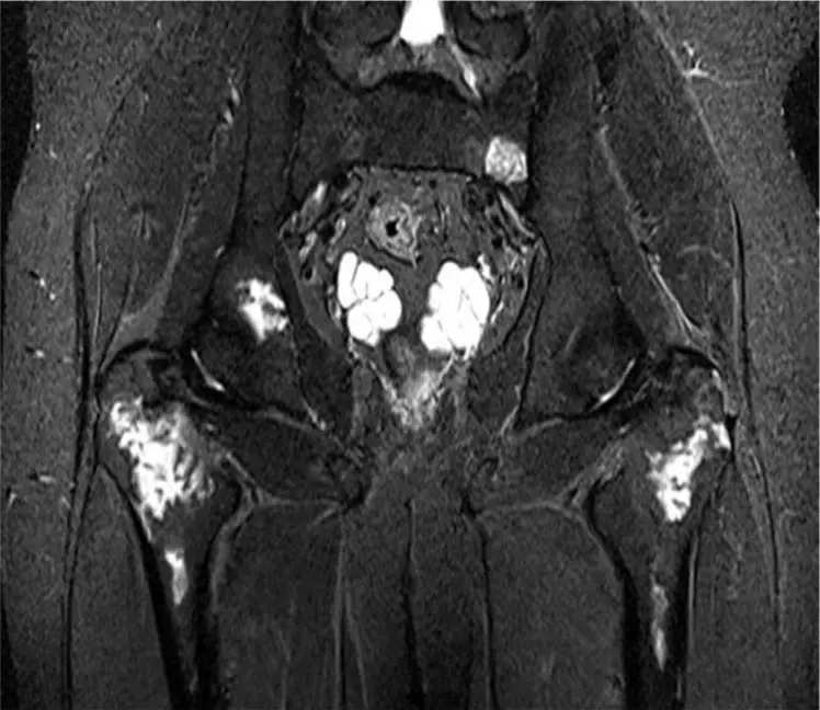
Infarcts, infection and marrow expansion: H-shaped vertebrae, collapsed femoral heads and Salmonella osteomyelitis.
Thalassemia, myelofibrosis and mastocytosis
Thalassemia expands the marrow until bones widen and sinuses disappear; myelofibrosis and mastocytosis make bones dense.
Differential: diffusely dense bones and the ivory vertebra
Ask the right questions in order: signs of hyperparathyroidism, the size of the spleen, axial predominance, fractures.
Differential: lucent epiphyseal lesions and expansile rib lesions
Two short lists worth memorizing: GEICHA for the epiphysis and FAMEB for an expanded rib.
Describing a fracture
A fracture report answers five questions: where, which direction, how many pieces, how displaced and angled, and does it reach the joint.
Stress, insufficiency, atypical and pathologic fractures
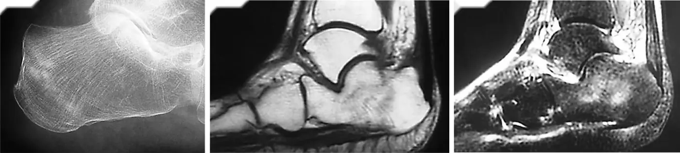
Abnormal stress on normal bone, normal stress on abnormal bone, a lateral femoral cortex after bisphosphonates, and a fracture through a lesion.
MRI of tendons: artifacts, tenosynovitis, tendinosis and tears
Normal tendons are black; gray can be real disease or the magic angle; fluid all the way around is tenosynovitis.
Forefoot overview and fifth metatarsal fractures
At the base of the fifth metatarsal, the zone decides the treatment: the tip is a simple avulsion, the junction is a Jones fracture that heals poorly.
Freiberg infraction and metatarsal stress fracture
A flattened second metatarsal head in a young woman, and a march fracture that shows only as faint periosteal new bone weeks after the pain starts.
Sesamoid fracture and turf toe
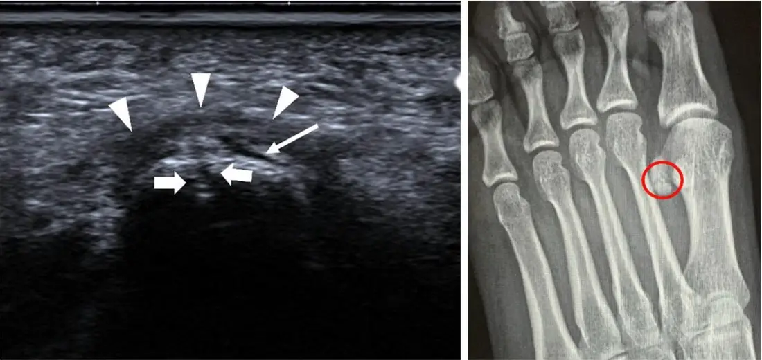
A split sesamoid with sharp, uncorticated edges is a fracture; smooth, rounded, corticated parts are a bipartite variant.
Further reading
- Ejindu VC, Hine AL, Mashayekhi M, et al. Musculoskeletal Manifestations of Sickle Cell Disease. RadioGraphics. 2007;27(4):1005-1021. doi:10.1148/rg.274065142
- Ihde LL, Forrester DM, Gottsegen CJ, et al. Sclerosing Bone Dysplasias: Review and Differentiation from Other Causes of Osteosclerosis. RadioGraphics. 2011;31(7):1865-1882. doi:10.1148/rg.317115093
- Pathria MN, Chung CB, Resnick DL. Acute and Stress-related Injuries of Bone and Cartilage: Pertinent Anatomy, Basic Biomechanics, and Imaging Perspective. Radiology. 2016;280(1):21-38. doi:10.1148/radiol.16142305
- Theodorou DJ, Theodorou SJ, Kakitsubata Y, et al. Fractures of Proximal Portion of Fifth Metatarsal Bone: Anatomic and Imaging Evidence of a Pathogenesis of Avulsion of the Plantar Aponeurosis and the Short Peroneal Muscle Tendon. Radiology. 2003;226(3):857-865. doi:10.1148/radiol.2263020284
- Hallinan JTPD, Statum SM, Huang BK, et al. High-Resolution MRI of the First Metatarsophalangeal Joint: Gross Anatomy and Injury Characterization. RadioGraphics. 2020;40(4):1107-1124. doi:10.1148/rg.2020190145
Read all 10 pages and answer the chapter's 30 questions in the app. Free with an account.