RadQBoard › Musculoskeletal › Chapter 2
Inflammatory and crystal arthropathies
The seronegative spondyloarthropathies share one mechanism, inflammation where tendons and ligaments attach to bone, and this chapter shows how it plays out in the sacroiliac joints, the spine and the small joints of the hands and feet. It then covers the connective tissue diseases, which deform and calcify more than they erode, and the three crystal diseases: CPPD, gout and hydroxyapatite deposition.
Read all 10 pages and answer the chapter's 34 questions in the app. Free with an account.
What this chapter covers
Seronegative spondyloarthropathies
One family, one mechanism: inflammation where tendons and ligaments attach to bone, which both erodes bone and makes new bone.
Sacroiliitis
Inflammation of the SI joint starts on the iliac side of the lower, synovial part of the joint, where the cartilage is thinnest.
Ankylosing spondylitis and IBD arthropathy
Advanced AS turns the spine into one rigid bone, and that rigid spine breaks easily.
Psoriatic and reactive arthritis
Psoriatic arthritis erodes and builds bone at the same time, mostly in the small joints of the hands, while bone density stays normal.
Lupus and dermatomyositis
The connective tissue diseases deform and calcify more than they erode: lupus bends the fingers without destroying bone, and dermatomyositis lays down sheets of calcium.
Scleroderma and acro-osteolysis
Scleroderma resorbs the fingertips and calcifies the soft tissues around them.
Calcium pyrophosphate deposition (CPPD)
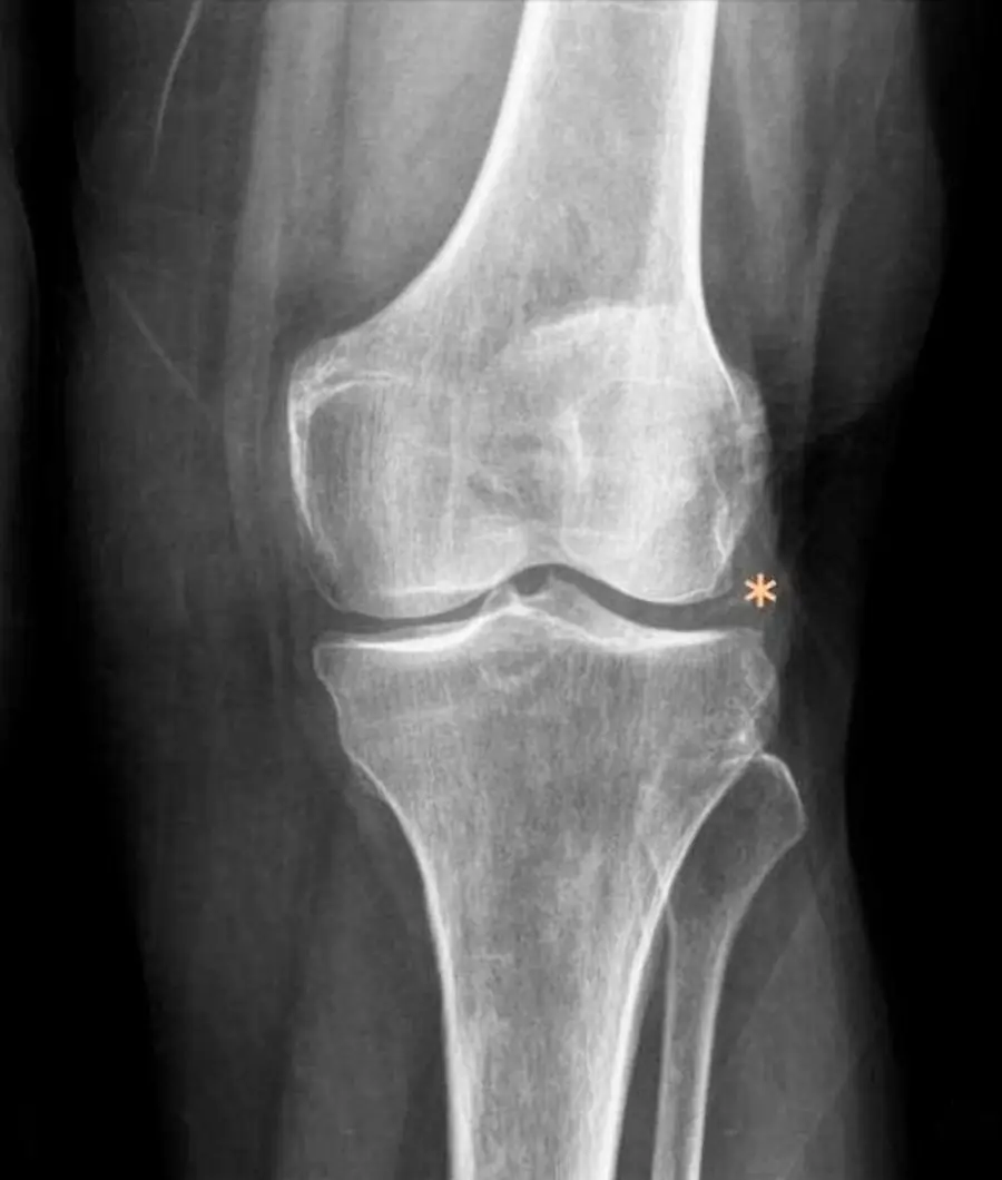
Calcium pyrophosphate settles inside cartilage and wears joints in unusual places.
GoutFree
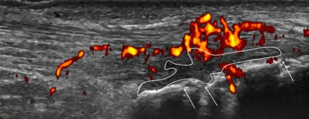
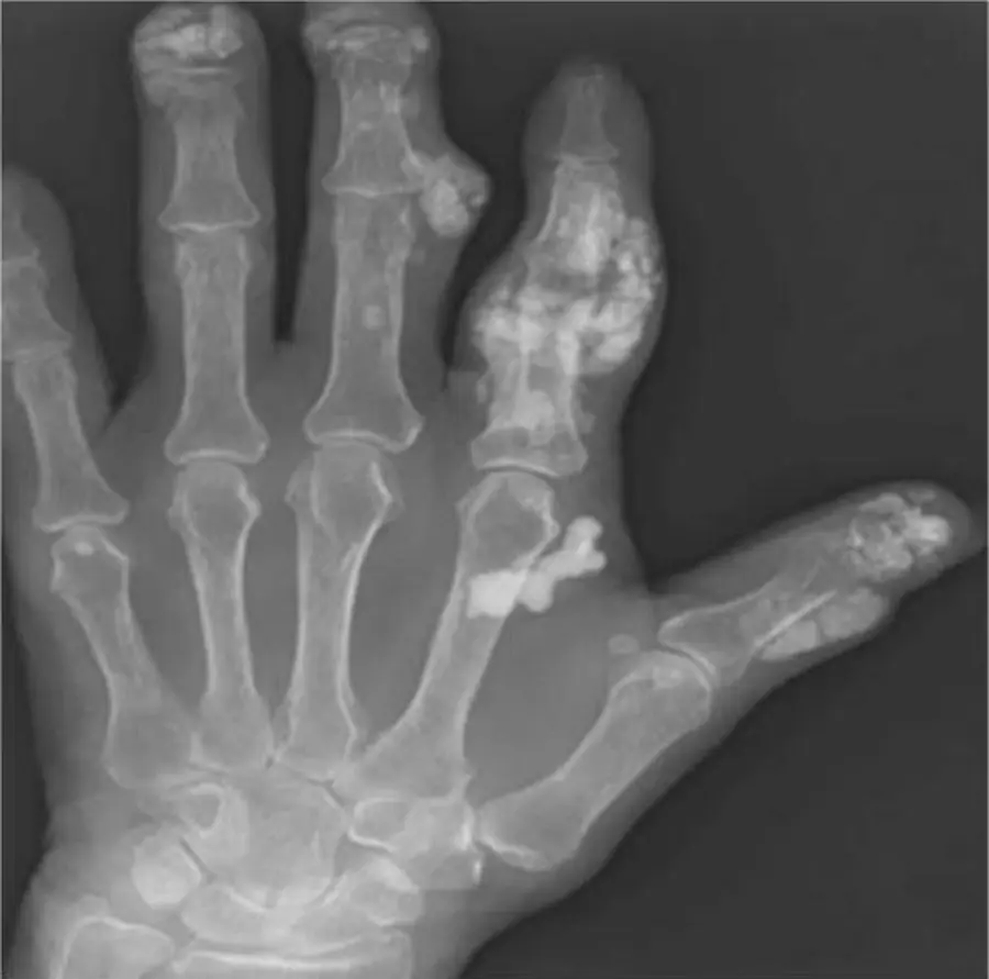
Urate builds tophi that punch holes in bone next to the joint while the joint space survives.
Hydroxyapatite deposition disease
Amorphous calcium in a tendon: no cortex, no trabeculae, and a tendency to look alarming on MRI.
Chapter review: pattern recognition
One table to test yourself: cover the right column and name the diagnosis.
Further reading
- Navallas M, Ares J, Beltrán B, et al. Sacroiliitis Associated with Axial Spondyloarthropathy: New Concepts and Latest Trends. RadioGraphics. 2013;33(4):933-956. doi:10.1148/rg.334125025
- Diekhoff T, Eshed I, Giraudo C, et al. Reporting Sacroiliac Joint Imaging Performed for Known or Suspected Axial Spondyloarthritis: Assessment of SpondyloArthritis International Society Recommendations. Radiology. 2024;311(3):e231786. doi:10.1148/radiol.231786
- Shiraishi M, Fukuda T, Igarashi T, et al. Differentiating Rheumatoid and Psoriatic Arthritis of the Hand: Multimodality Imaging Characteristics. RadioGraphics. 2020;40(5):1339-1354. doi:10.1148/rg.2020200029
- Freire V, Moser TP, Lepage-Saucier M. Radiological identification and analysis of soft tissue musculoskeletal calcifications. Insights into Imaging. 2018;9(4):477-492. doi:10.1007/s13244-018-0619-0 · Free full text
- Aswani Y, Nasirul Islam GM, Scholz FJ, et al. Imaging in Systemic Sclerosis (Scleroderma). RadioGraphics. 2026;46(12):e250193. doi:10.1148/rg.250193
- Steinbach LS, Resnick D. Calcium pyrophosphate dihydrate crystal deposition disease revisited. Radiology. 1996;200(1):1-9. doi:10.1148/radiology.200.1.8657894
- Desai MA, Peterson JJ, Garner HW, et al. Clinical Utility of Dual-Energy CT for Evaluation of Tophaceous Gout. RadioGraphics. 2011;31(5):1365-1375. doi:10.1148/rg.315115510
Read all 10 pages and answer the chapter's 34 questions in the app. Free with an account.