RadQBoard › Musculoskeletal › Chapter 2 › Gout
Gout
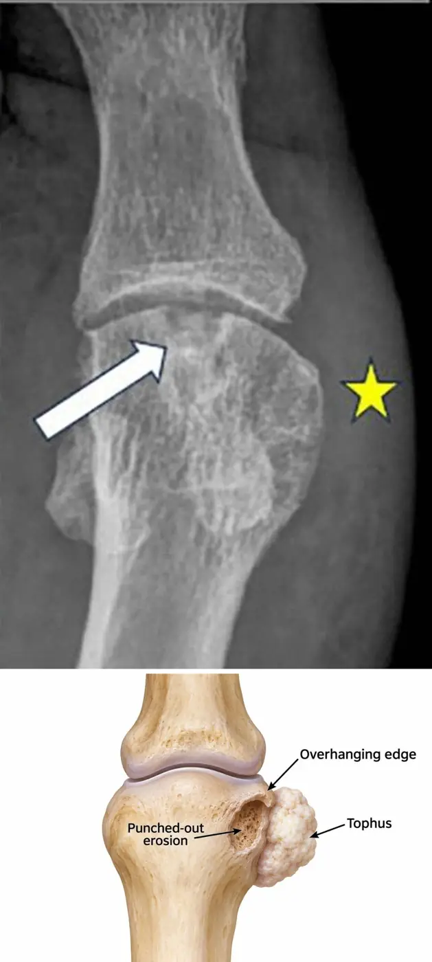
Urate builds tophi that punch holes in bone next to the joint while the joint space survives.
What it is
Gout is deposition of monosodium urate crystals in and around joints, after years of high serum urate.
- Mostly middle-aged men and postmenopausal women.
- Acute attacks are red, hot and very painful; chronic tophaceous gout follows years of attacks.
- Risk factors include renal disease, diuretics, alcohol and a purine-rich diet.
Where it strikes
First MTP joint most often (podagra), then midfoot, ankle, knee, olecranon bursa and hands.
Radiographs
Radiographs change only after years:
- Well-defined, punched-out erosions with sclerotic rims and overhanging edges, often away from the joint margin.
- Joint space and bone density preserved until late.
- Tophi: dense, lumpy soft-tissue masses that may calcify.
Ultrasound
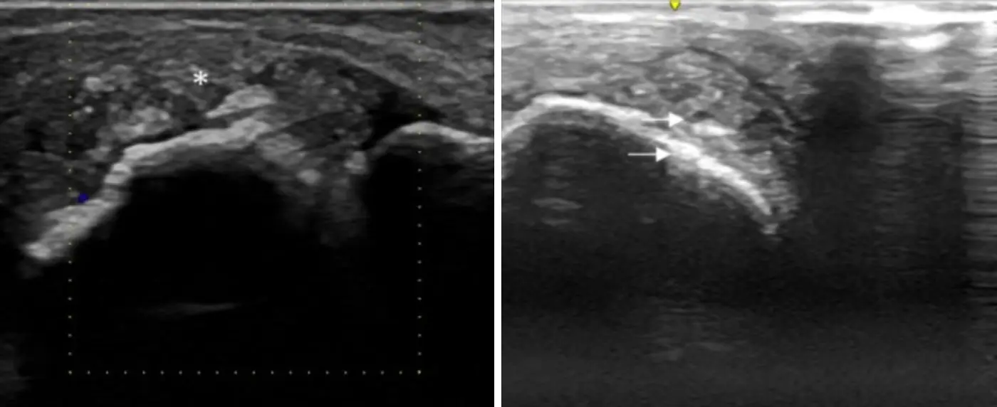
- Double contour sign: a bright irregular line of urate on the surface of the hyaline cartilage, parallel to the bone.
- Tophi: bright aggregates with a dark rim.
- CPPD can mimic it (pseudo-double contour): CPP sits inside the cartilage, urate on its surface.
Dual-energy CT
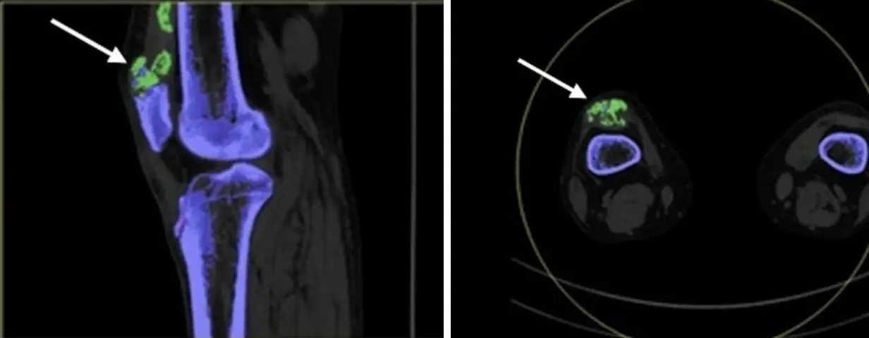
- Material decomposition labels urate deposits; the software chooses the color, often green.
- False coding can come from skin, nail beds, metal and beam hardening.
- It supports the diagnosis and measures urate burden, but can be negative in early or low-volume gout.
- Crystals on imaging do not exclude a coexisting septic joint.
Differential diagnosis
| Feature | Gout | Rheumatoid arthritis |
|---|---|---|
| Erosions | Punched out, sclerotic rim, overhanging edge, often away from the margin | Marginal, ill-defined |
| Joint space | Preserved until late | Lost early, uniformly |
| Bone density | Normal | Periarticular osteopenia |
| Soft tissue | Lumpy, asymmetric tophi | Symmetric fusiform swelling |
- A tophus can mimic a tumor or infection on MRI: usually low to intermediate on T1, heterogeneous on T2, with enhancement. Check the radiograph and DECT.
Further reading
- Desai MA, Peterson JJ, Garner HW, et al. Clinical Utility of Dual-Energy CT for Evaluation of Tophaceous Gout. RadioGraphics. 2011;31(5):1365-1375. doi:10.1148/rg.315115510
Practice this in RadQBoard. Chapter 2 has 34 board-style questions with full explanations. Free with an account.
Written in original words by the RadQBoard team. Images are reused under open licenses, with the source, license and any changes under each one.