RadQBoard › Musculoskeletal › Chapter 1 › Rheumatoid arthritis of the cervical spine
Rheumatoid arthritis of the cervical spine
RA erodes and loosens the neck without building any bone; the danger point is C1–C2.
What it is
The cervical spine is involved in up to 70% of patients, more with severe, long-standing disease. Synovitis of the atlantoaxial joints and the synovial joints around the dens loosens the ligaments that hold C1 and C2 together.
The general pattern
- Subluxation at several levels, often a stepladder appearance below C2.
- Osteopenia.
- Erosions of the dens, facets, endplates and spinous processes (which can taper to points).
- No bone production, unlike OA and the spondyloarthropathies.
The transverse ligament
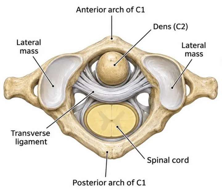
The transverse ligament runs behind the dens between the lateral masses of C1 and holds the dens against the anterior arch of C1. Inflammation and laxity of this ligament allow C1 to slide forward on C2.
Atlantoaxial subluxation
| Direction | Cause | Imaging |
|---|---|---|
| Anterior (most common) | Laxity of the transverse ligament | Atlanto-dental interval (ADI) > 2.5 mm in adults (> 5 mm in children), often only on flexion views |
| Vertical (atlantoaxial impaction, cranial settling) | Erosion and collapse of the C1–C2 facets | Dens rises through the foramen magnum toward the brainstem; the anterior arch of C1 sinks to the level of the C2 body |
| Posterior | Erosion or fracture of the dens | C1 slides back on C2 |
| Rotatory and lateral | Asymmetric facet destruction | Open-mouth view, CT |
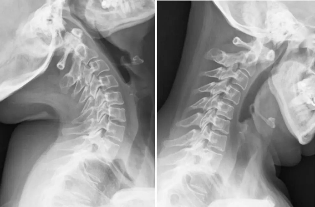
The ADI is measured on a lateral radiograph from the back of the anterior arch of C1 to the front of the dens. Anterior subluxation may not show without a flexion view. The space left for the cord behind the dens (the posterior atlanto-dental interval) matters more for neurologic risk: a small posterior interval warns of cord compression.
- A posterior atlantodental interval (dens to the back of the ring of C1) under about 14 mm predicts cord compression better than the anterior interval.
- Cranial settling (basilar invagination): the dens tip more than about 4.5 mm above McGregor's line can compress the brainstem.
Imaging
- Lateral radiographs in neutral, flexion and extension for instability.
- The dens tip is often hidden on radiographs; in vertical subluxation the anterior arch of C1 sinking to the C2 body is the clue.
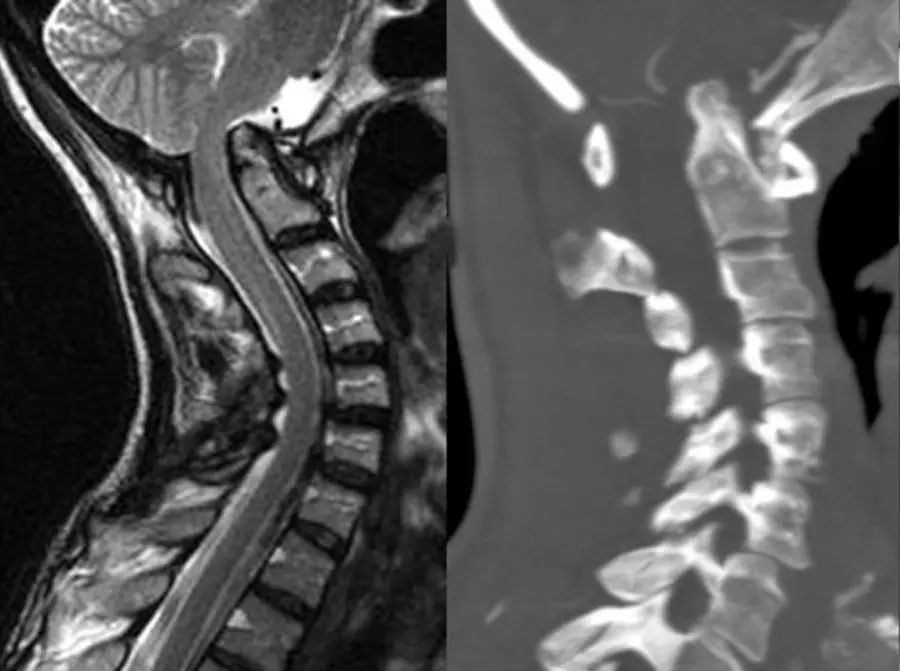
- CT shows erosions and alignment.
- MRI shows pannus around the dens and compression of the cord or brainstem.
Clinical relevance
- Neck pain, myelopathy or brainstem symptoms; sudden death is possible in severe impaction.
- Before intubation or surgery, patients with long-standing RA often need cervical spine imaging, because neck flexion can injure the cord.
Further reading
- Morimoto LR, Kase DT, Esmanhotto PG, et al. Imaging Assessment of Nontraumatic Pathologic Conditions at the Craniovertebral Junction: A Comprehensive Review. RadioGraphics. 2024;44(5):e230137. doi:10.1148/rg.230137
Practice this in RadQBoard. Chapter 1 has 32 board-style questions with full explanations. Free to read, no account needed.
Written in original words by the RadQBoard team. Images are reused under open licenses, with the source, license and any changes under each one.