RadQBoard › Musculoskeletal › Chapter 1 › Rheumatoid arthritis of the foot, hip and knee
Rheumatoid arthritis of the foot, hip and knee Musculoskeletal · Chapter 1: Bones, joints, osteoarthritis and rheumatoid arthritis · Page 9 of 10 · Free to read
In the foot RA can be the first sign; in the hip and knee it narrows the whole joint evenly.
Foot
The MTP joints of the forefoot and the talocalcaneonavicular joint of the midfoot are typical.
Up to 1 in 5 patients present with an MTP joint as the first site, so foot radiographs belong in the work-up even without foot symptoms.
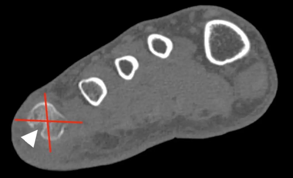
The lateral side of the 5th metatarsal head is a classic site of early erosion.
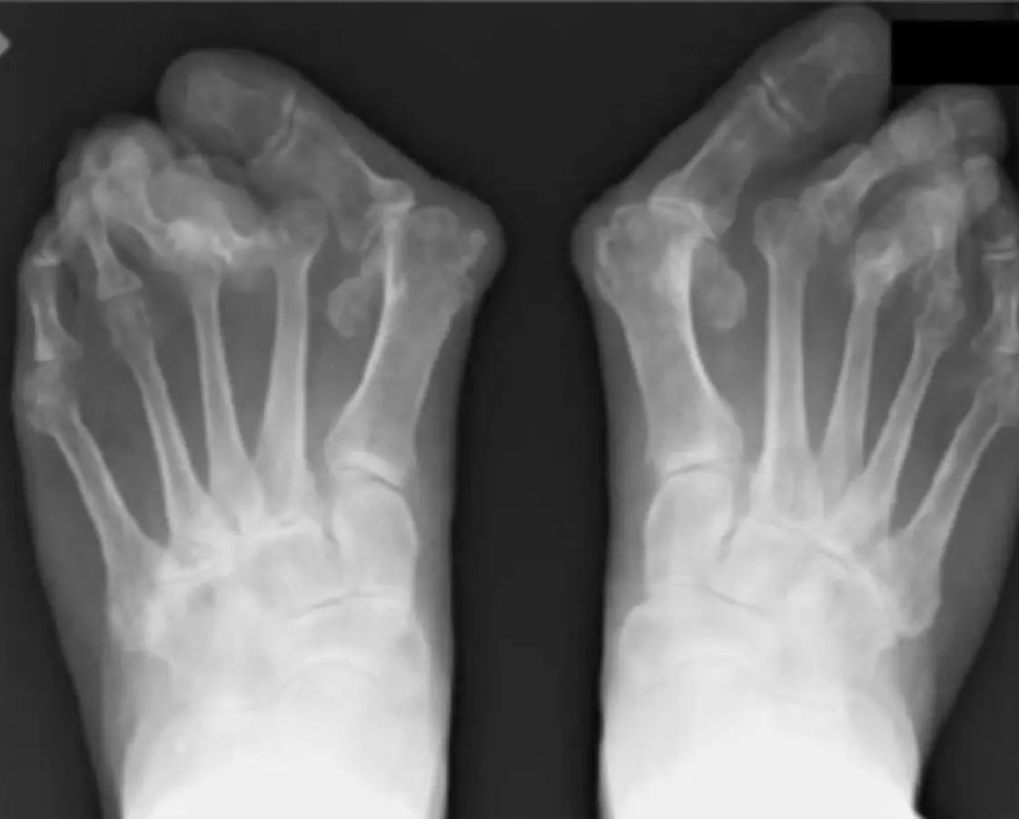
Later: hallux valgus, lateral (fibular) deviation of the toes and subluxed MTP joints.
At the heel: retrocalcaneal bursitis with erosion of the posterior calcaneus.
The classic early site in the foot. CT through the metatarsal heads of the right foot: an erosion at the lateral (fibular) quadrant of the fifth metatarsal head (arrowhead); the red lines divide the head into quadrants. Image: Simoni P, Moussaddykine S, Malaise O, et al. Topography of bone erosions at the metatarsophalangeal joints in rheumatoid arthritis: bilateral mapping by computed tomography. Cureus 2021;13:e15823 (Fig. 2A) . CC BY 4.0 . Changes: cropped (right foot of panel A); marks from the original figure. Rheumatoid forefoot in a 75-year-old woman with 40 years of RA (weight-bearing view): severe hallux valgus, subluxation and dislocation of the lesser MTP joints with fibular drift of the toes, and joint erosions. Image: Etani Y, Hirao M, Ebina K, et al. Combination of modified scarf osteotomy and metatarsal shortening offset osteotomy for rheumatoid forefoot deformity. Int J Environ Res Public Health 2021;18:10473 (Fig. 1a) . CC BY 4.0 . Changes: cropped to the preoperative panel; panel letter and a dark label box removed; converted to grayscale. Hip
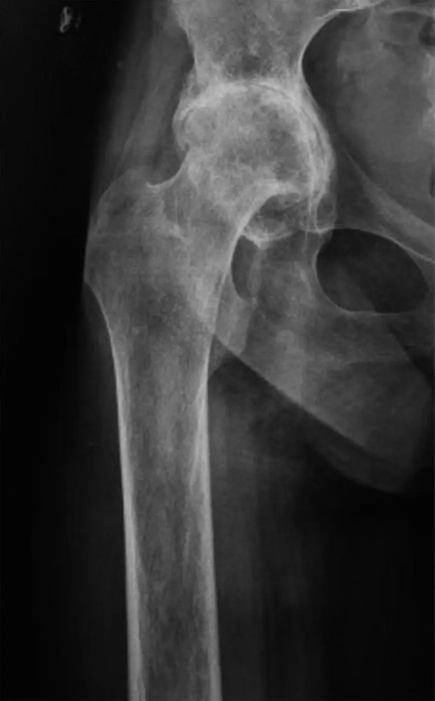
Concentric (axial) cartilage loss moves the femoral head straight in along the neck axis, unlike the superolateral migration of OA.
Severe cases develop protrusio acetabuli : the femoral head crosses the ilioischial line by more than 3 mm in men or 6 mm in women.
Osteophytes are small or absent unless secondary OA has developed.
Rheumatoid hip in a 36-year-old woman: concentric loss of the joint space with the femoral head pushed deep into the acetabulum, no osteophytes, severe osteoporosis and a wide femoral canal with thin cortex. Image: Zhen P, Chang Y, Yue H, et al. Primary total hip arthroplasty using a short bone-conserving stem in young adult osteoporotic patients with Dorr type C femoral bone. J Orthop Surg Res 2021;16:17 (Fig. 3a) . CC BY 4.0 . Changes: cropped; panel letter and side marker removed. Knee
All three compartments (medial and lateral tibiofemoral, patellofemoral) narrow evenly, whereas OA starts in the medial tibiofemoral compartment.
Erosions are less prominent than in the small joints.
Osteophytes with uniform narrowing mean secondary OA on top of RA.
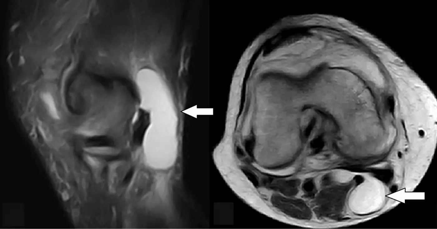
Large effusions and popliteal (Baker) cysts are common; a ruptured cyst can mimic deep vein thrombosis.
Popliteal (Baker) cyst on MRI, sagittal (left) and axial (right): fluid distends the gastrocnemius–semimembranosus bursa behind the knee (arrows) and communicates with the joint through a posterior capsular opening. Large cysts are common with rheumatoid effusions, and rupture can mimic deep vein thrombosis. Image: Zmerly H, Di Lorenzo L, Mahfouz V, et al. Ruptured Baker's cyst demystified: current evidence, diagnostic strategies, and treatment options for an under-recognized condition. Cureus 2026;18:e101686 (Fig. 1) . CC BY 4.0 . Changes: cropped; panel letters removed; arrows from the original figure; converted to grayscale. Shoulder
Erosion of the superolateral humeral head and tapering of the distal clavicle.
Rotator cuff thinning and tears with a high-riding humeral head.
In the shoulder: erosions at the greater tuberosity, a high-riding humeral head as the cuff thins, and an eroded, tapered distal clavicle.
Key points
Image the feet in suspected RA: the MTP joints can be the first site.
Hip: axial migration and protrusio.
Knee: even narrowing of all three compartments; osteophytes mean secondary OA.
Further reading Martínez de Vega L, Baker J, Suevos-Ballesteros C, et al. A Radiologic Walk through Rheumatoid Arthritis. RadioGraphics. 2026;46(3):e250059. doi:10.1148/rg.250059 ← Rheumatoid arthritis of the hand and wrist Rheumatoid arthritis of the cervical spine → Written in original words by the RadQBoard team. Images are reused under open licenses, with the source, license and any changes under each one.