RadQBoard › Musculoskeletal › Chapter 1 › Rheumatoid arthritis of the hand and wrist
Rheumatoid arthritis of the hand and wrist
MCP, PIP and wrist, both sides, with the DIP joints spared: the rheumatoid hand.
Where it strikes
- MCP joints, PIP joints and the carpal joints, symmetrically.
- The DIP joints are usually spared.
Early radiographs
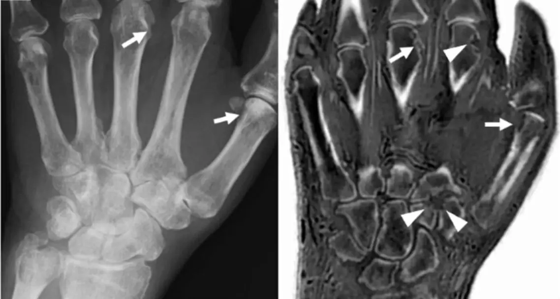
- Fusiform soft-tissue swelling and periarticular osteopenia, from synovitis and hyperemia.
- Early erosions at typical spots:
- the radial side of the 2nd and 3rd metacarpal heads;
- the radial and ulnar sides of the bases of the proximal phalanges;
-
the ulnar styloid.
-
Early erosions are often at the ulnar styloid, the second and third MCP heads, and in the feet at the fifth MTP joint. Image the feet even when only the hands hurt.
Late radiographs: deformities
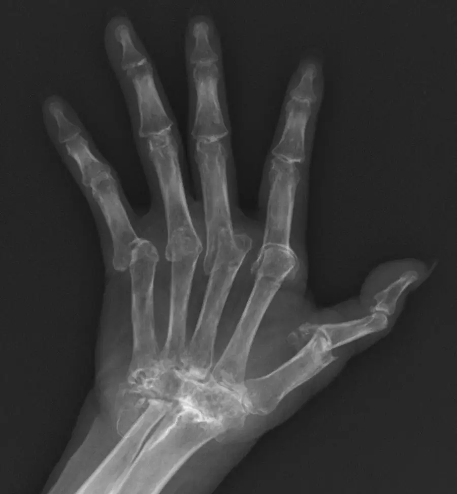
Advanced RA subluxates joints, and the deformities are usually fixed (not reducible), unlike lupus:
- Ulnar deviation of the fingers at the MCP joints.
- Swan neck: PIP hyperextension with DIP flexion.
- Boutonnière: PIP flexion with DIP hyperextension.
- Z-shaped thumb.
The wrist
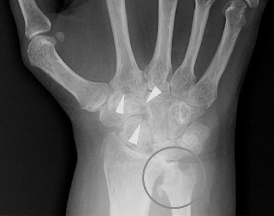
- Narrowing and erosions across the radiocarpal, intercarpal and carpometacarpal joints.
- Carpal collapse and ulnar translocation of the carpus; scapholunate widening.
- Erosion of the ulnar styloid and distal ulna, which can rupture the extensor tendons of the little and ring fingers.
- Late-stage RA can uncommonly fuse the wrist (ankylosis after complete cartilage loss). Carpal ankylosis is much more typical of juvenile idiopathic arthritis.
Differential diagnosis
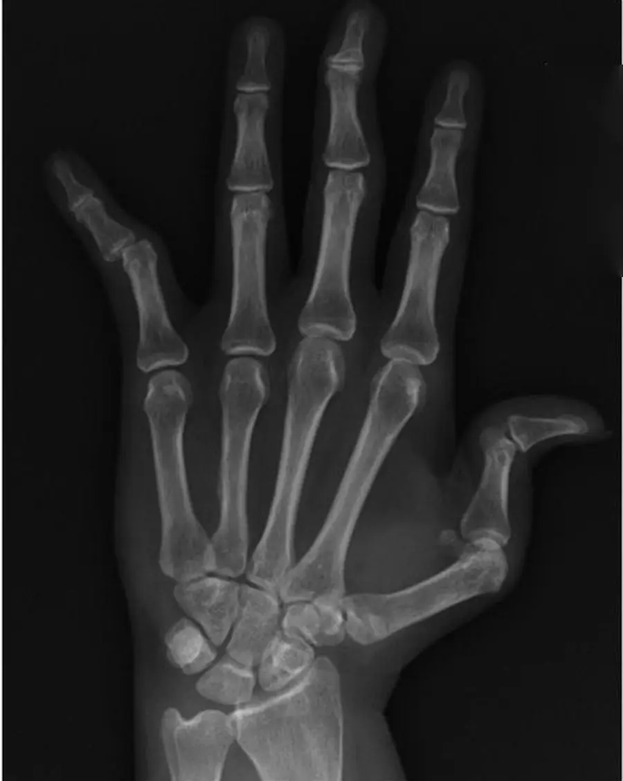
| Feature | Rheumatoid arthritis | Lupus (Jaccoud) | Psoriatic arthritis | Gout |
|---|---|---|---|---|
| Erosions | Marginal | None | Marginal, pencil-in-cup | Punched out, away from the joint |
| Deformities | Fixed | Reducible | Telescoping digits late | Lumpy tophi |
| Density | Osteopenic | Osteopenic or normal | Normal | Normal |
| Joints | MCP, PIP, wrist | MCP | DIP, PIP, ray | Any, asymmetric |
Further reading
- Sommer O, Kladosek A, Weiler V, et al. Rheumatoid Arthritis: A Practical Guide to State-of-the-Art Imaging, Image Interpretation, and Clinical Implications. RadioGraphics. 2005;25(2):381-398. doi:10.1148/rg.252045111
Practice this in RadQBoard. Chapter 1 has 32 board-style questions with full explanations. Free to read, no account needed.
Written in original words by the RadQBoard team. Images are reused under open licenses, with the source, license and any changes under each one.