RadQBoard › Musculoskeletal › Chapter 1 › Rheumatoid arthritis: the disease
Rheumatoid arthritis: the disease
Rheumatoid arthritis is an attack on the synovium; the bone loss is collateral damage, starting where synovium touches bare bone.
What it is
Rheumatoid arthritis (RA) is an autoimmune disease in which the synovium is the target of a waxing and waning immune attack.
- More often women, usually from middle age.
- Symmetric joint pain and swelling with morning stiffness.
- Rheumatoid factor (an antibody against IgG that activates complement) is usually positive but not specific; anti-CCP antibodies are more specific.
Where it strikes
- First the small joints of the hands and wrists. The feet are often involved early, so foot radiographs are part of the routine work-up.
- Later the cervical spine, knees, shoulders and hips.
- The DIP joints are usually spared.
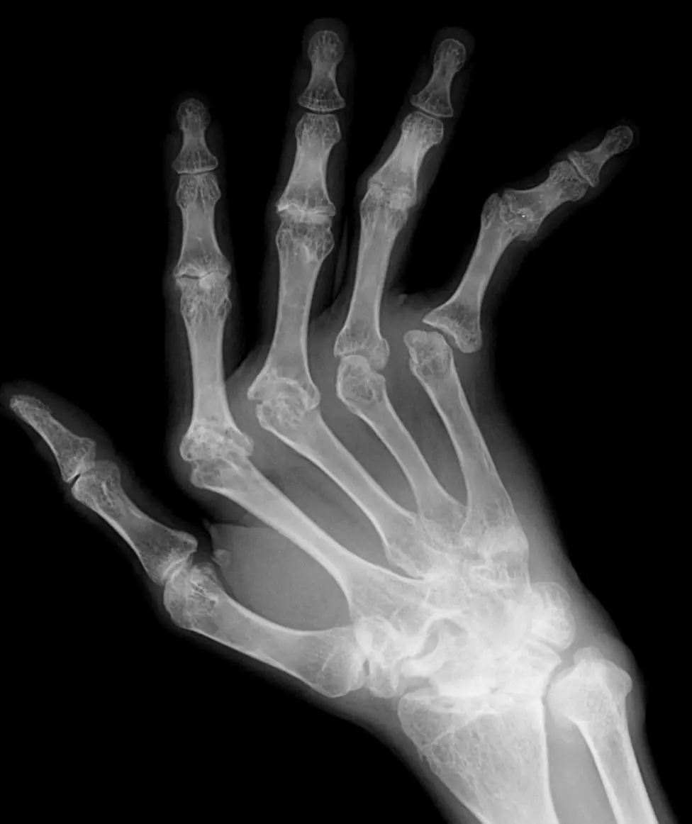
Why the bone erodes
Erosions are the imaging and pathologic hallmark of RA: focal bone destruction from cumulative inflammation. Two mechanisms are proposed: inflamed synovium (pannus) eating into bone from outside ("outside-in") and inflammation inside the bone ("inside-out"). About half of patients have erosions after one year of symptoms, and erosions predict disability and worse quality of life, which is why early detection and treatment matter.
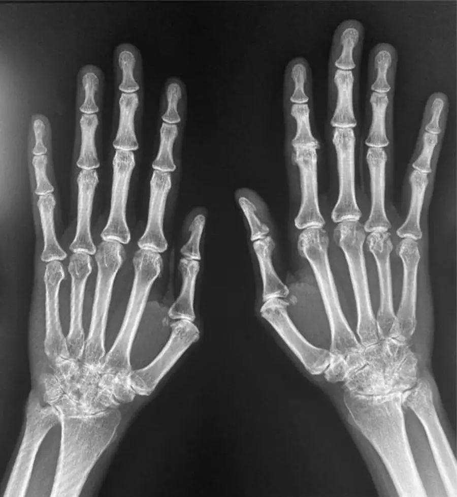
Radiographs and CT
- Soft-tissue swelling around the joints.
- Periarticular osteopenia, one of the earliest signs.
- Marginal erosions, first at the bare area inside the capsule, where bone is not covered by cartilage.
- Uniform, symmetric joint-space narrowing.
- Subluxations and deformities later.
- Little or no new bone; osteophytes mean secondary OA.
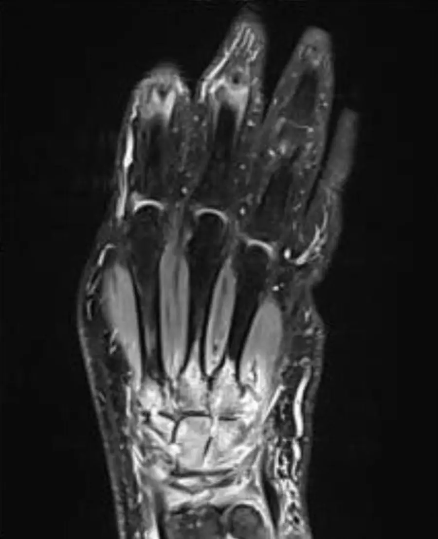
MRI and ultrasound
- Synovitis: thickened synovium that enhances early after contrast.
- Erosions: sharply marginated defects with fluid signal that enhance.
- Bone marrow edema (osteitis): subchondral edema from inflammation; it often comes before erosions.
- Tenosynovitis and joint subluxation.
- Ultrasound shows synovitis with Doppler flow and small erosions, and guides treatment decisions.
- In early RA, bone marrow edema on MRI is the strongest predictor of future erosions.
Differential diagnosis
| Feature | Rheumatoid arthritis | Osteoarthritis |
|---|---|---|
| Joints | MCP, PIP, wrist, MTP | DIP, PIP, 1st CMC, knee, hip |
| Narrowing | Uniform | Uneven |
| Density | Periarticular osteopenia | Normal |
| Erosions | Marginal | None (central in erosive OA) |
| New bone | None | Osteophytes, sclerosis |
Further reading
- Sommer O, Kladosek A, Weiler V, et al. Rheumatoid Arthritis: A Practical Guide to State-of-the-Art Imaging, Image Interpretation, and Clinical Implications. RadioGraphics. 2005;25(2):381-398. doi:10.1148/rg.252045111
- Martínez de Vega L, Baker J, Suevos-Ballesteros C, et al. A Radiologic Walk through Rheumatoid Arthritis. RadioGraphics. 2026;46(3):e250059. doi:10.1148/rg.250059
Practice this in RadQBoard. Chapter 1 has 32 board-style questions with full explanations. Free to read, no account needed.
Written in original words by the RadQBoard team. Images are reused under open licenses, with the source, license and any changes under each one.