RadQBoard › Musculoskeletal › Chapter 1 › Degenerative spine and DISH
Degenerative spine and DISH
The disc degenerates; the facets get osteoarthritis. They are two different processes in two different kinds of joint.
What it is
- The disc–vertebral body junction is a cartilaginous joint: the annulus fibrosus and nucleus pulposus (the disc) and the hyaline cartilage endplates. Its wear is called discovertebral degeneration, more accurate than "degenerative disc disease"; it is best seen as a chronic response to injury.
- The spine's synovial joints get true osteoarthritis: facet (zygapophyseal), atlantoaxial, uncovertebral (C3–C7), costovertebral and sacroiliac joints.
Discovertebral degeneration: imaging
- Disc desiccation (dark on T2) and loss of height.
- Endplate sclerosis and remodeling; on MRI, endplate signal changes (Modic type 1 edema, type 2 fat, type 3 sclerosis).
- Osteophytes from the vertebral body margins.
- Vacuum phenomenon: gas (nitrogen) inside the disc. It is a sign of degeneration and argues strongly against infection.
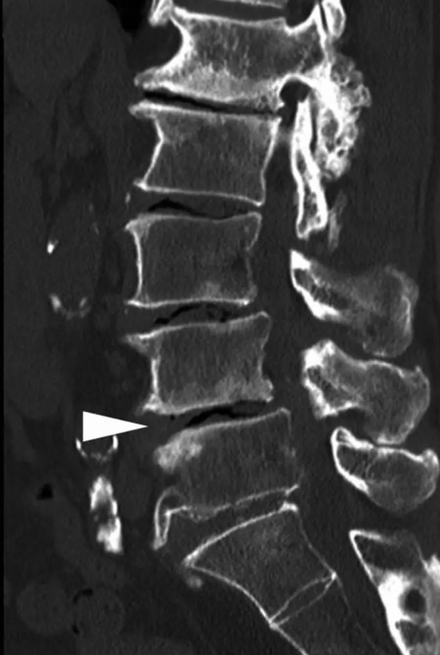
Vacuum disc or Kümmell disease?
Do not confuse gas in the disc with gas in the vertebral body. Kümmell disease is gas (a vacuum cleft) inside a collapsed vertebral body after a compression fracture, from osteonecrosis and non-union. The cleft can fill with fluid on MRI, and the fracture can keep collapsing.
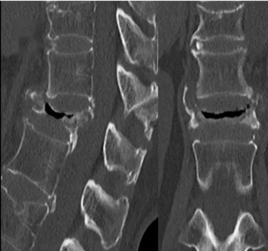
Complications
- Central spinal canal stenosis.
- Neural foraminal stenosis.
- Degenerative spondylolisthesis, most often at L4–L5, from facet OA with an intact pars.
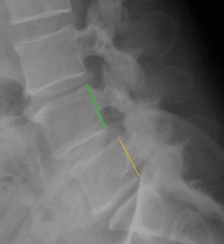
DISH: what it is
Diffuse idiopathic skeletal hyperostosis (DISH) is ossification of ligaments and entheses of unknown cause in older adults, more often men, associated with obesity and diabetes. It looks like exuberant osteophytes but is a separate disease.
DISH: imaging
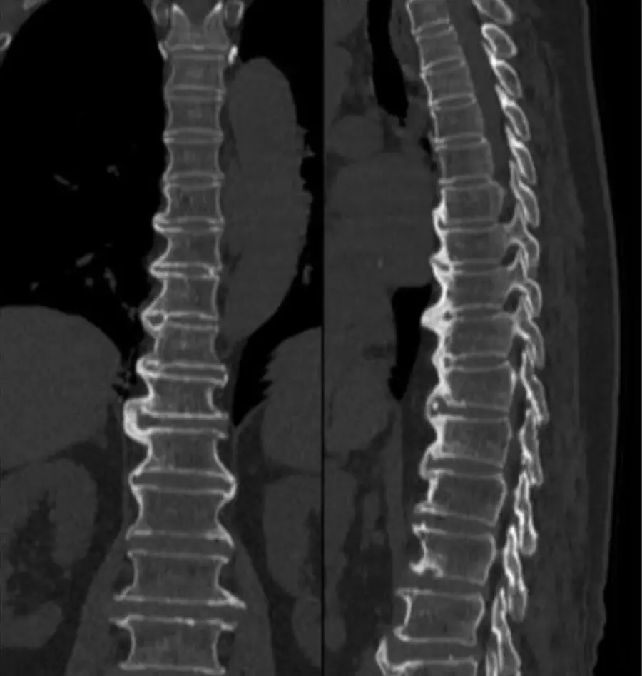
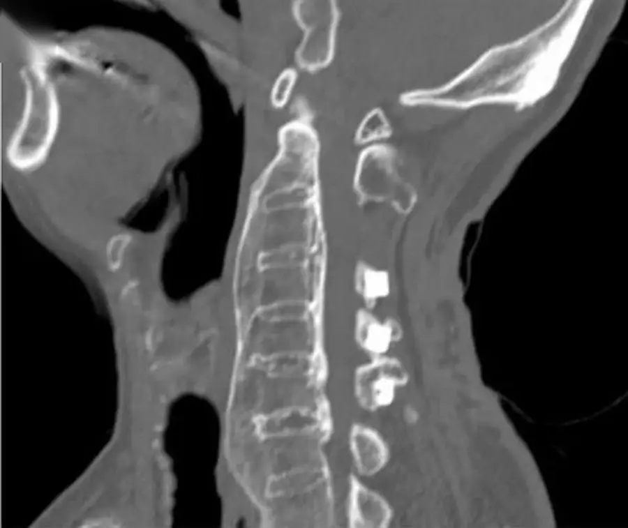
- Flowing, bridging ossification along the front and side of the spine across at least four contiguous vertebral bodies.
- Normal disc heights, and no erosions or fusion of the synovial SI joints; facets are not fused.
- Most common in the thoracic spine, typically on the right side, away from the pulsating aorta.
- Enthesophytes elsewhere: pelvis, heels, patella, olecranon.
- Usually silent; in the cervical spine large anterior bridges can cause dysphagia.
- Associated with ossification of the posterior longitudinal ligament (OPLL), which can narrow the canal; OPLL can be hard to see on MRI and is best shown by CT.
- A spine stiffened by DISH breaks like a long bone after minor trauma, as in ankylosing spondylitis.
- OPLL (ossification of the posterior longitudinal ligament) often comes with DISH and can narrow the cervical canal; CT shows it best.
Differential diagnosis
| Feature | DISH | Ankylosing spondylitis | Degenerative osteophytes |
|---|---|---|---|
| Age | Older | Young adult | Older |
| Bridging bone | Thick, flowing, anterior and right-sided, ≥4 levels | Thin, marginal syndesmophytes | Horizontal spurs that may bridge |
| Discs | Preserved | Preserved | Narrowed, vacuum |
| SI joints | Normal synovial part | Erosions, fusion | Normal or OA |
| Facets | Open | Fused | OA |
Further reading
- Modic MT, Ross JS. Lumbar Degenerative Disk Disease. Radiology. 2007;245(1):43-61. doi:10.1148/radiol.2451051706
- Resnick D, Niwayama G. Radiographic and Pathologic Features of Spinal Involvement in Diffuse Idiopathic Skeletal Hyperostosis (DISH). Radiology. 1976;119(3):559-568. doi:10.1148/119.3.559
Practice this in RadQBoard. Chapter 1 has 32 board-style questions with full explanations. Free to read, no account needed.
Written in original words by the RadQBoard team. Images are reused under open licenses, with the source, license and any changes under each one.