RadQBoard › Musculoskeletal › Chapter 1 › Osteoarthritis of the hip
Osteoarthritis of the hip
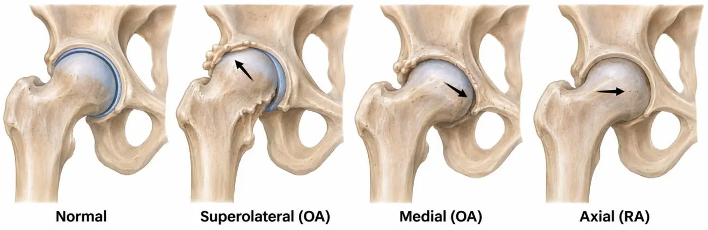
The direction the femoral head moves tells you the arthritis: up and out in OA, straight in along the axis in rheumatoid arthritis.
What it is
Hip OA is cartilage loss of the hip joint, usually bilateral. Much of it is secondary to an abnormal shape of the joint: femoroacetabular impingement (cam or pincer), developmental dysplasia, previous slipped capital femoral epiphysis or Perthes disease, osteonecrosis, or trauma.
Patterns of narrowing and migration
| Pattern | Femoral head moves | Typical of |
|---|---|---|
| Superolateral | Up and out | Osteoarthritis, the most common pattern |
| Medial | Inward and down | Osteoarthritis, less common |
| Axial (concentric) | Straight in, along the femoral neck axis | Rheumatoid and other inflammatory arthritis |
Axial narrowing is the typical pattern of rheumatoid arthritis, but OA is far more common. So axial narrowing with osteophytes is still more likely OA; axial narrowing without osteophytes favors rheumatoid arthritis.
Radiographs
- Narrowing in one of the patterns above.
- Osteophytes around the femoral head and acetabular rim.
- Subchondral sclerosis and cysts on both sides of the joint.
- Buttressing: thickening of the medial cortex of the femoral neck.
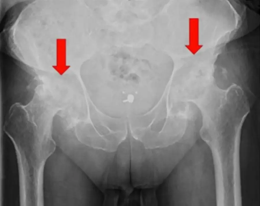
Protrusio acetabuli
The femoral head pushes medially past the inner wall of the pelvis.
- Defined on a frontal radiograph when the femoral head crosses the ilioischial line by more than 3 mm in men or 6 mm in women.
- Causes: rheumatoid arthritis and other inflammatory arthritis, bone softening (osteomalacia, Paget disease, hyperparathyroidism), Marfan syndrome, trauma, and an idiopathic form.
- On the AP pelvis, protrusio is present when the femoral head crosses the ilioischial line by more than about 3 mm in men or 6 mm in women.
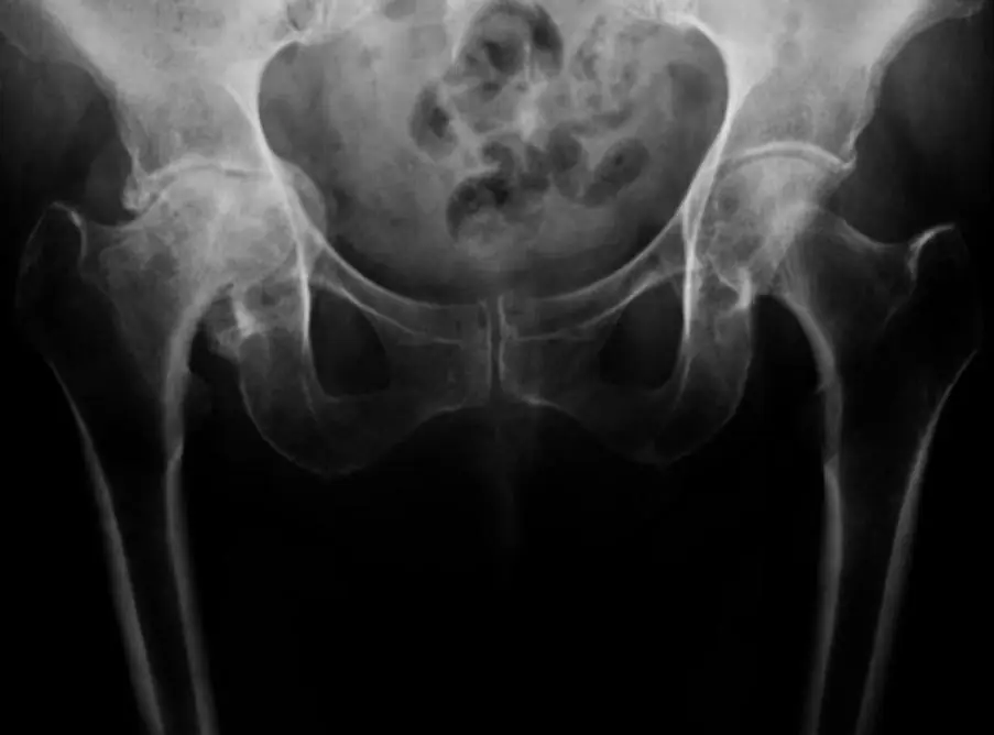
Differential diagnosis
- Rapidly destructive hip OA: an older patient, usually a woman, loses the cartilage and part of the femoral head within months. It mimics infection, osteonecrosis and neuropathic joint; aspiration may be needed to exclude infection.
- Septic arthritis: rapid loss of the joint space with erosions and effusion in an ill patient.
Further reading
- Modic MT, Ross JS. Lumbar Degenerative Disk Disease. Radiology. 2007;245(1):43-61. doi:10.1148/radiol.2451051706
Practice this in RadQBoard. Chapter 1 has 32 board-style questions with full explanations. Free to read, no account needed.
Written in original words by the RadQBoard team. Images are reused under open licenses, with the source, license and any changes under each one.