RadQBoard › Musculoskeletal › Chapter 1 › Osteoarthritis of the knee, shoulder and foot
Osteoarthritis of the knee, shoulder and foot
Weight-bearing views tell the truth about the knee; the Grashey view tells it about the shoulder.
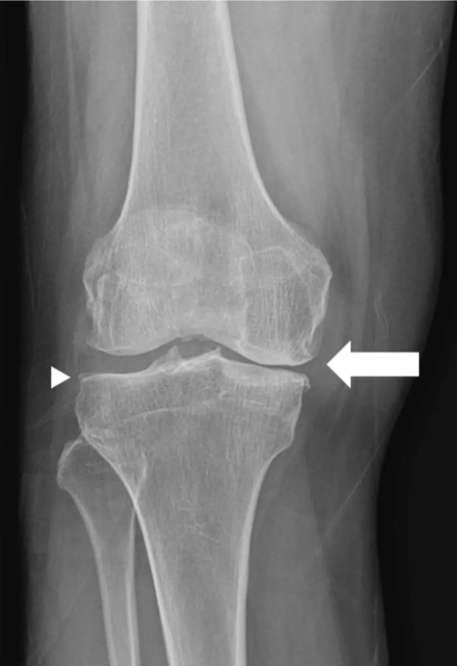
Knee: where it strikes
- The knee has three compartments: medial tibiofemoral, lateral tibiofemoral and patellofemoral.
- OA typically starts in the medial tibiofemoral compartment, asymmetrically; severe OA involves all three.
- Both knees are usually involved.
- Isolated or dominant patellofemoral disease suggests CPPD; uniform narrowing of all compartments without osteophytes suggests an inflammatory arthritis.
Knee: imaging
- Standing views show the true cartilage space; a non-weight-bearing film can look normal. A standing PA view in slight flexion shows the posterior cartilage, which wears first, and is more sensitive than a standing AP view in extension.
- Osteophytes decide that OA is present; narrowing grades its severity.
- Medial narrowing tips the knee into varus.
- MRI shows cartilage loss, subchondral marrow edema-like signal, meniscal tears and extrusion, and osteophytes.
- Severe patellofemoral narrowing out of proportion to the other compartments, especially with chondrocalcinosis, suggests CPPD.
Shoulder
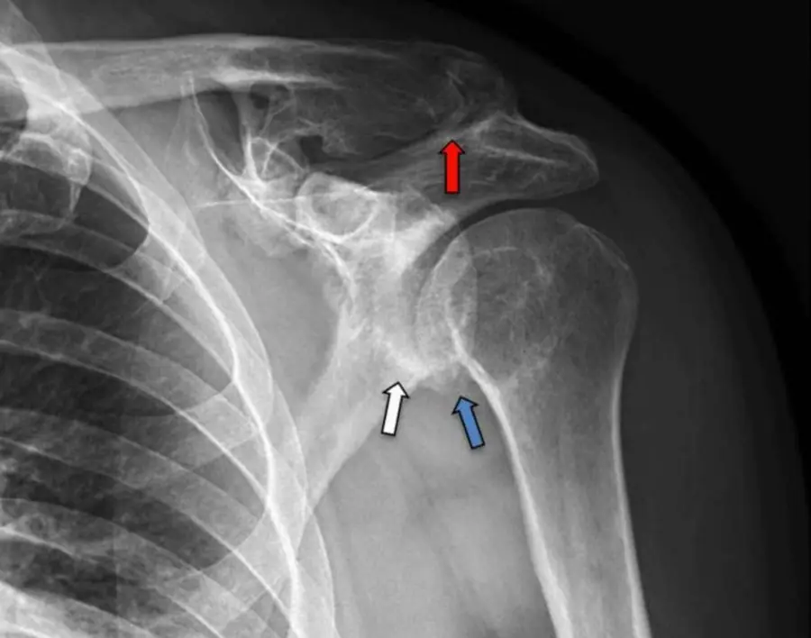
- The Grashey view (a 40° posterior oblique view in external rotation) puts the glenohumeral joint in profile and shows its narrowing best.
- Primary glenohumeral OA is uncommon. Look for a cause: prior instability or surgery, rotator cuff tear arthropathy, CPPD, osteonecrosis or trauma.
- A ring of osteophyte on the inferior humeral head is the typical early sign; the glenoid wears posteriorly.
- Glenohumeral OA is unusual without a cause: look for old trauma, instability surgery, CPPD or a chronic cuff tear.
Foot
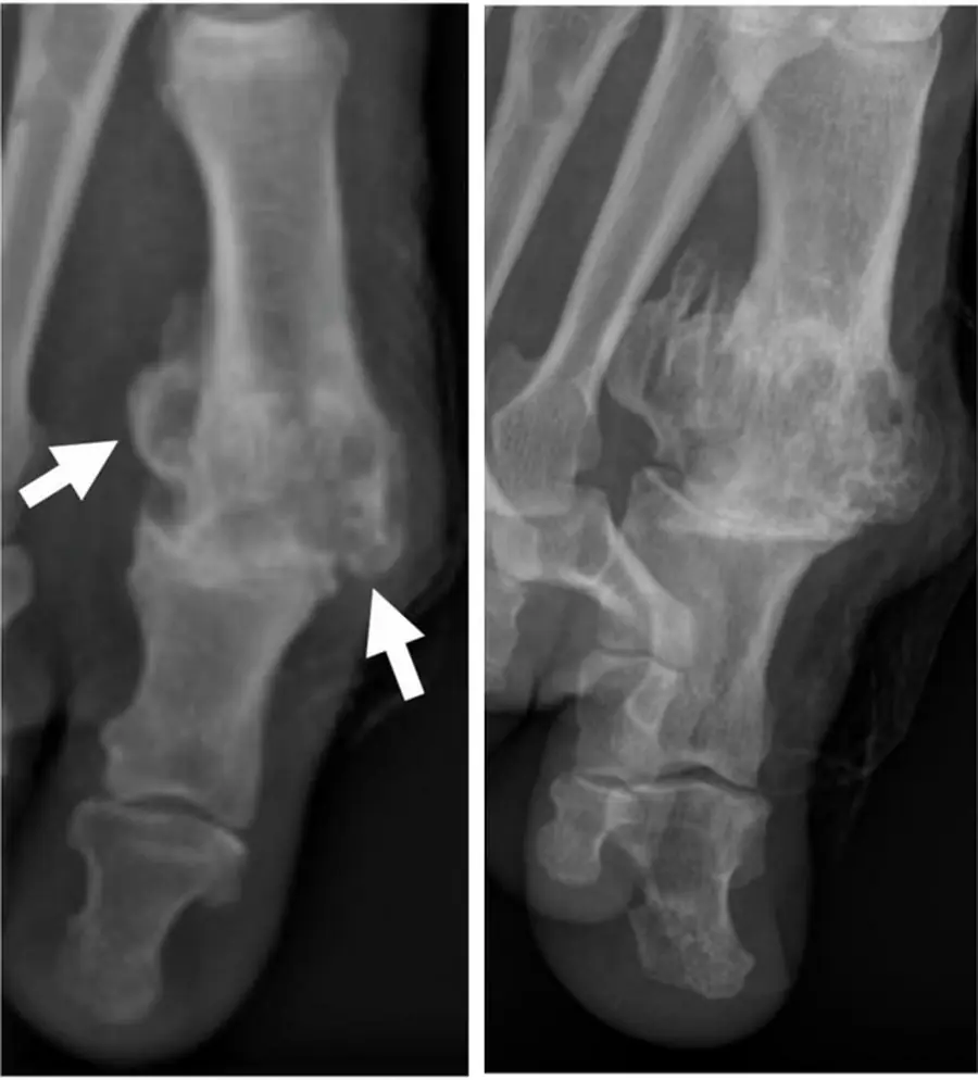
- First MTP joint: the most common site of foot OA. Dorsal osteophytes and narrowing stiffen the joint, called hallux rigidus.
- Talonavicular joint: OA causes dorsal beaking at the joint margin. Do not confuse it with the talar beak of a tarsal coalition, which arises from the dorsal talar head in a young patient.
- Ankle OA is usually post-traumatic.
Further reading
- Roemer FW, Demehri S, Omoumi P, et al. State of the Art: Imaging of Osteoarthritis—Revisited 2020. Radiology. 2020;296(1):5-21. doi:10.1148/radiol.2020192498
Practice this in RadQBoard. Chapter 1 has 32 board-style questions with full explanations. Free to read, no account needed.
Written in original words by the RadQBoard team. Images are reused under open licenses, with the source, license and any changes under each one.