RadQBoard › Musculoskeletal › Chapter 1 › Osteoarthritis of the hand and erosive OA
Osteoarthritis of the hand and erosive OA
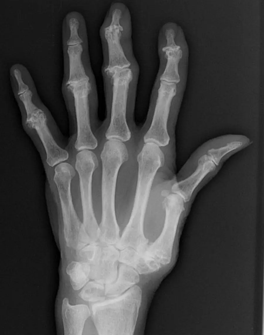
In the hand, OA lives at the DIP joints, the thumb base and the PIP joints, and leaves the MCP joints alone.
Where it strikes
In order of frequency:
- DIP joints.
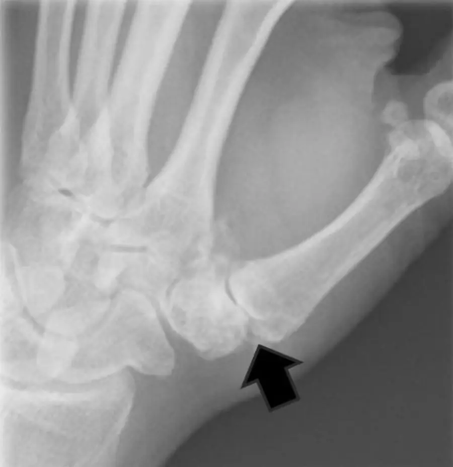
- First carpometacarpal (CMC) joint, the thumb base, often with the scaphotrapeziotrapezoid (STT) joint.
- PIP joints.
The MCP joints are usually spared, which separates OA from rheumatoid arthritis. MCP osteophytes, especially at the 2nd and 3rd joints, point to CPPD or hemochromatosis.
Radiographs
- Narrowing, subchondral sclerosis and osteophytes; no erosions.
- Heberden nodes: firm swelling around the DIP joints from osteophytes.
- Bouchard nodes: the same at the PIP joints.
- At the thumb base: narrowing and osteophytes of the first CMC joint, often with radial subluxation of the first metacarpal base.
- Apart from the first CMC joint, primary OA in the wrist involves the STT (scaphotrapeziotrapezoid) joint. OA of the radiocarpal joint alone suggests old trauma or CPPD.
Erosive osteoarthritis: what it is
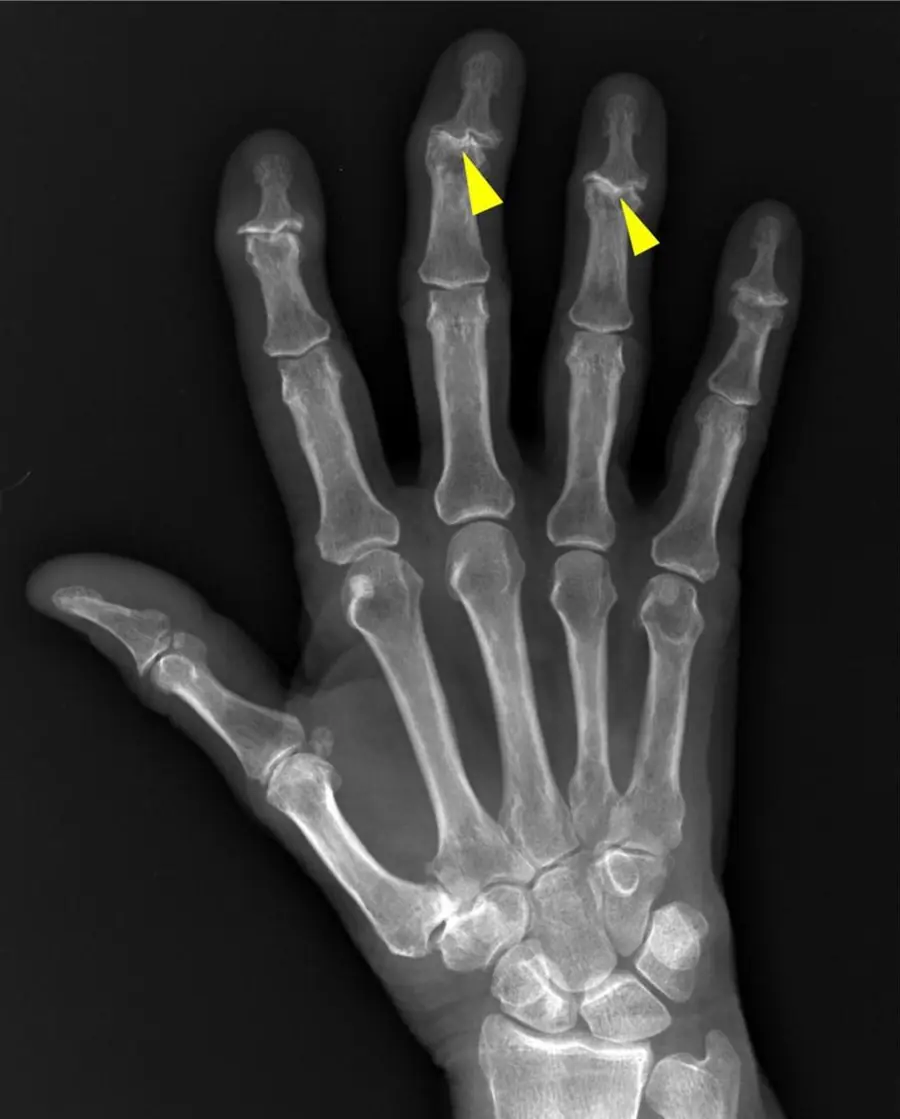
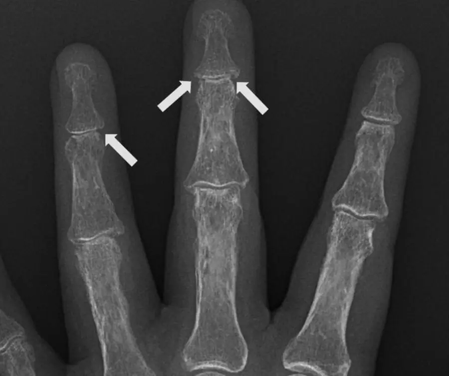
Erosive OA is an inflammatory form of hand OA, typically in older women. Clinically it behaves like an inflammatory arthritis (pain, swelling, redness, stiffness), but its distribution and most of its imaging look like OA.
Erosive OA: imaging
- Limited to the hands: DIP and PIP joints, symmetric.
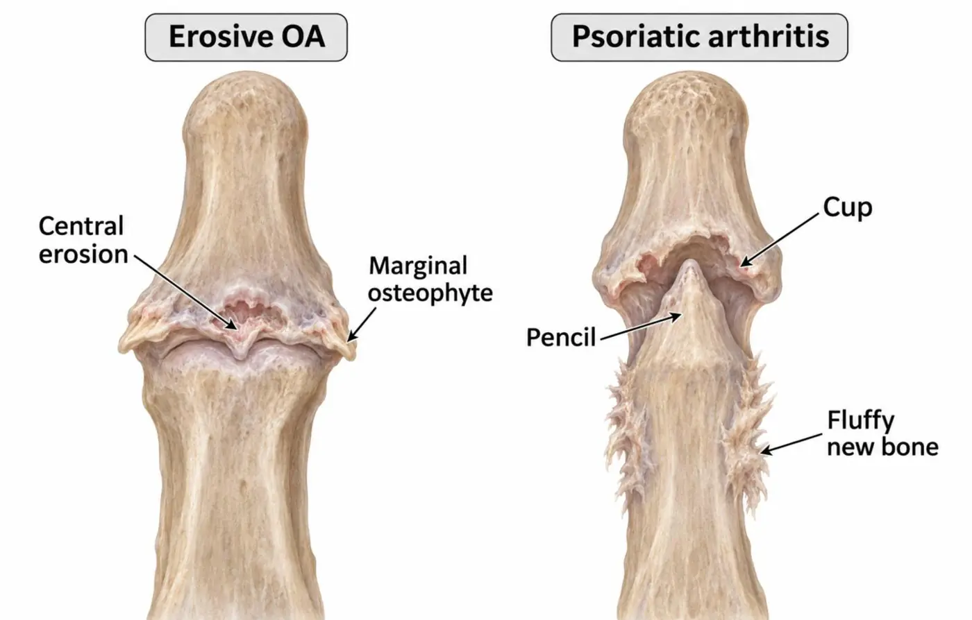
- Central erosions with marginal osteophytes. Together they make the gull-wing shape of the joint: the eroded, thinned center with osteophytes spreading to each side, like the wings of a gull in flight.
- Bone density stays normal; there is no periostitis.
- The interphalangeal joints can fuse late in the disease.
Differential diagnosis
| Feature | Erosive OA | Psoriatic arthritis | Rheumatoid arthritis |
|---|---|---|---|
| Joints | DIP, PIP | DIP, PIP, a whole ray | MCP, PIP, wrist |
| Erosions | Central (gull-wing) | Marginal, then pencil-in-cup | Marginal |
| New bone | Osteophytes | Fluffy periostitis | None |
| Density | Normal | Normal | Periarticular osteopenia |
| Symmetry | Symmetric | Often asymmetric | Symmetric |
Further reading
- Martel W, Stuck KJ, Dworin AM, et al. Erosive osteoarthritis and psoriatic arthritis: a radiologic comparison in the hand, wrist, and foot. American Journal of Roentgenology. 1980;134(1):125-135. doi:10.2214/ajr.134.1.125
Practice this in RadQBoard. Chapter 1 has 32 board-style questions with full explanations. Free to read, no account needed.
Written in original words by the RadQBoard team. Images are reused under open licenses, with the source, license and any changes under each one.