RadQBoard › Musculoskeletal › Chapter 1 › Osteoarthritis: the disease
Osteoarthritis: the disease
Osteoarthritis wears cartilage away where the load falls, and the bone answers with osteophytes, sclerosis and cysts.
What it is
Osteoarthritis (OA) is breakdown of articular cartilage from abnormal local mechanics in a susceptible person. It is a disease of the whole joint: bone, ligaments, menisci, capsule, synovium and muscle all take part.
- The most common cause of cartilage loss in middle-aged and older adults.
- Primary OA follows the typical distribution with no underlying cause.
- Secondary OA follows something else: trauma, a previous arthritis, joint malformation (hip dysplasia, femoroacetabular impingement), osteonecrosis or crystal disease.
Where it strikes
- Weight-bearing joints: knees (medial compartment), hips (superolateral), spine (facets) and the first MTP joint.
- Hands: DIP joints, the first carpometacarpal joint and PIP joints.
- OA in a young patient or an unusual joint (shoulder, elbow, wrist, ankle, MCP joints) usually means prior trauma or another arthritis, such as CPPD or hemochromatosis.
Radiographs and CT
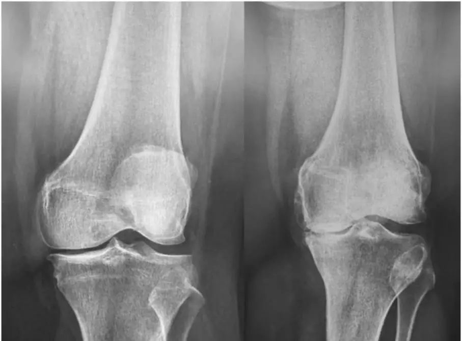
- Joint-space narrowing, usually uneven: worst where the load is greatest.
- Osteophytes: bony spurs at the joint margins. Osteophytes decide whether OA is present; narrowing decides how severe it is.
- Subchondral sclerosis from reactive remodeling.
- Subchondral cysts (geodes), from joint fluid forced into bone through cartilage defects.
- No erosions and no periarticular osteopenia.
OA can be diagnosed with confidence when narrowing comes with osteophytes, sclerosis and subchondral cysts, and erosions and periarticular osteopenia are absent.
Kellgren–Lawrence grade (most used for the knee):
| Grade | Findings |
|---|---|
| 0 | Normal |
| 1 | Doubtful narrowing, possible tiny osteophyte |
| 2 | Definite osteophytes, possible narrowing |
| 3 | Multiple osteophytes, definite narrowing, some sclerosis |
| 4 | Large osteophytes, marked narrowing, severe sclerosis, deformity |
MRI
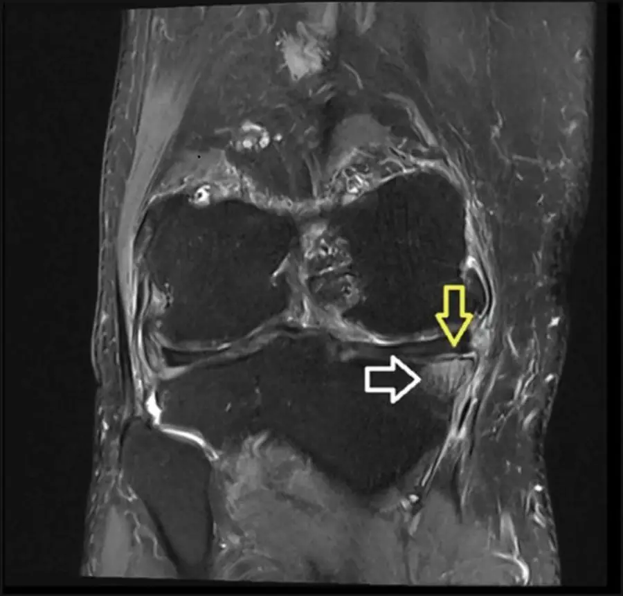
- Cartilage thinning, fissures and full-thickness defects.
- Osteophytes and subchondral cysts.
- Subchondral bone marrow edema-like signal, from inflammatory and reactive change; it tracks with pain.
- Synovitis: thick, irregular, enhancing synovium.
- In the knee, meniscal degeneration, tears and extrusion.
- Bone marrow lesions (edema-like subchondral signal) in osteoarthritis go with pain and predict faster cartilage loss.
Differential diagnosis
| Feature | Osteoarthritis | Rheumatoid arthritis | CPPD arthropathy |
|---|---|---|---|
| Narrowing | Uneven, loaded part first | Uniform | Uneven, unusual joints |
| Density | Normal | Periarticular osteopenia | Normal |
| Osteophytes | Yes | No (unless secondary OA) | Yes, often large |
| Erosions | No | Marginal | No |
| Clue | Typical joints | Symmetric small joints | Chondrocalcinosis, patellofemoral, radiocarpal, 2nd–3rd MCP |
Further reading
- Roemer FW, Demehri S, Omoumi P, et al. State of the Art: Imaging of Osteoarthritis—Revisited 2020. Radiology. 2020;296(1):5-21. doi:10.1148/radiol.2020192498
Practice this in RadQBoard. Chapter 1 has 32 board-style questions with full explanations. Free to read, no account needed.
Written in original words by the RadQBoard team. Images are reused under open licenses, with the source, license and any changes under each one.